Review
. 2020 Mar 21;9(3):234.
doi: 10.3390/pathogens9030234.

The Bradyzoite: A Key Developmental Stage for the Persistence and Pathogenesis of Toxoplasmosis

Affiliations
Review

The Bradyzoite: A Key Developmental Stage for the Persistence and Pathogenesis of Toxoplasmosis

Aude Cerutti et al. Pathogens. .

Abstract

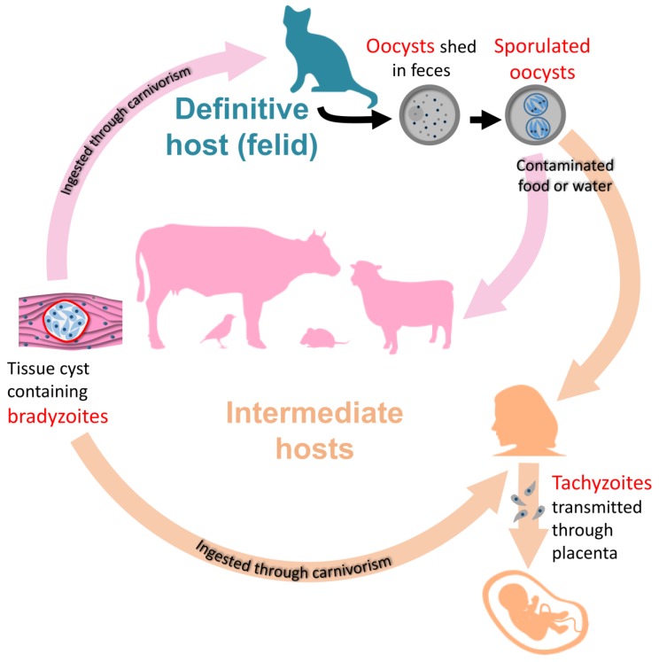
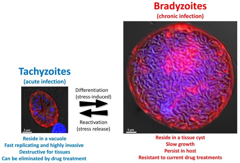
Toxoplasma gondii is a ubiquitous parasitic protist found in a wide variety of hosts, including a large proportion of the human population. Beyond an acute phase which is generally self-limited in immunocompetent individuals, the ability of the parasite to persist as a dormant stage, called bradyzoite, is an important aspect of toxoplasmosis. Not only is this stage not eliminated by current treatments, but it can also reactivate in immunocompromised hosts, leading to a potentially fatal outcome. Yet, despite its critical role in the pathology, the bradyzoite stage is relatively understudied. One main explanation is that it is a considerably challenging model, which essentially has to be derived from in vivo sources. However, recent progress on genetic manipulation and in vitro differentiation models now offers interesting perspectives for tackling key biological questions related to this particularly important developmental stage.

Keywords: Toxoplasma gondii; bradyzoite; chronic toxoplasmosis; differentiation; latency; metabolism; persistence.


Conflict of interest statement

The authors declare no conflict of interest. The funders had no role in the in the writing of in the decision to publish the manuscript.

Figures

Figure 1
Figure 1
The life cycle of Toxoplasma gondii. Bradyzoites occupy a central place in the transmission of the parasite, between the intermediate, or to the definitive hosts.
Figure 2
Figure 2
T. gondii can be found in intermediate hosts as two different stages called tachyzoite and bradyzoite. Images represent an in vitro grown tachyzoite-containing vacuole (left) and a bradyzoite-containing tissue cyst extracted from a mouse brain (right). The parasitophorous vacuole membrane was labelled with an anti-GRA3 antibody (red, left), the cyst wall was labelled with Dolichos biflorus agglutinin (red, right). DNA was labelled with 4′,6-diamidino-2-phenylindole (DAPI, blue). Images represent merged fluorescence and differential interference contrast (DIC) micrographs.
Figure 3
Figure 3
Spontaneous differentiation of bradyzoites in neurons and myotubes. Cysts (arrowhead, labelled with Dolichos biflorus agglutinin, green) form spontaneously in primary hippocampal neurons from mouse (left, labelled with anti- Microtubule Associated Protein 2, red) or immortalized human myotubes (right, labelled with anti-myosin heavy chain, red). DNA was labelled with with 4′,6-diamidino-2-phenylindole (DAPI, blue).

References

    1. Dubey J.P. Toxoplasma Gondii. Elsevier; Amsterdam, The Netherlands: 2014. The history and life cycle of Toxoplasma gondii; pp. 1–17.
    1. Shapiro K., Bahia-Oliveira L., Dixon B., Dumètre A., de Wit L.A., VanWormer E., Villena I. Environmental transmission of Toxoplasma gondii: Oocysts in water, soil and food. Food Waterborne Parasitol. 2019;15:e00049. doi: 10.1016/j.fawpar.2019.e00049. - DOI - PMC - PubMed
    1. Montoya J.G., Liesenfeld O. Toxoplasmosis. Lancet. 2004;363:1965–1976. doi: 10.1016/S0140-6736(04)16412-X. - DOI - PubMed
    1. McAuley J.B. Congenital Toxoplasmosis. J. Pediatr. Infect. Dis. Soc. 2014;3:S30–S35. doi: 10.1093/jpids/piu077. - DOI - PMC - PubMed
    1. Remington J.S., Cavanaugh E.N. Isolation of the encysted form of Toxoplasma gondii from human skeletal muscle and brain. N. Engl. J. Med. 1965;273:1308–1310. doi: 10.1056/NEJM196512092732404. - DOI - PubMed

LinkOut - more resources