. 2020 Mar 21;12(3):744.
doi: 10.3390/cancers12030744.

Antibody-Drug Conjugate Using Ionized Cys-Linker-MMAE as the Potent Payload Shows Optimal Therapeutic Safety

Affiliations

Antibody-Drug Conjugate Using Ionized Cys-Linker-MMAE as the Potent Payload Shows Optimal Therapeutic Safety

Yanming Wang et al. Cancers (Basel). .

Abstract

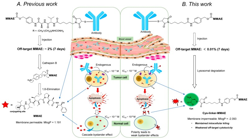
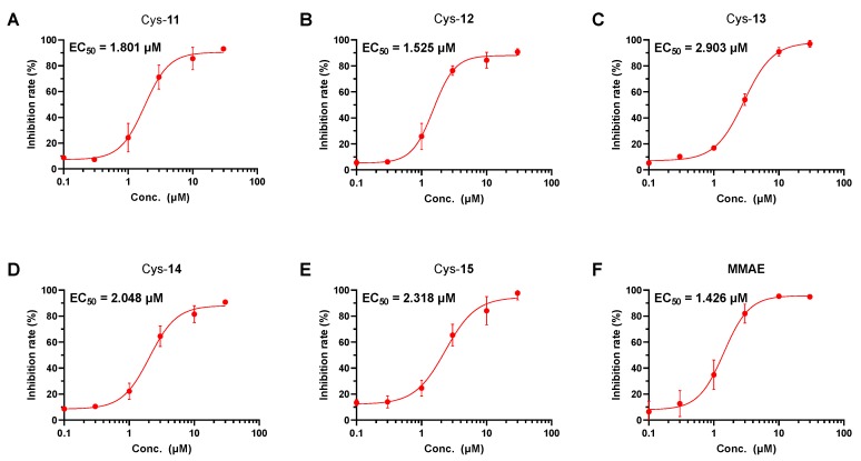
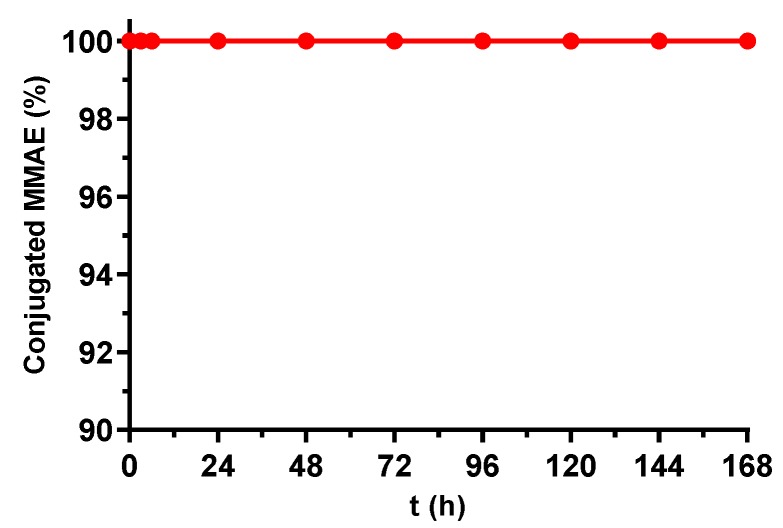
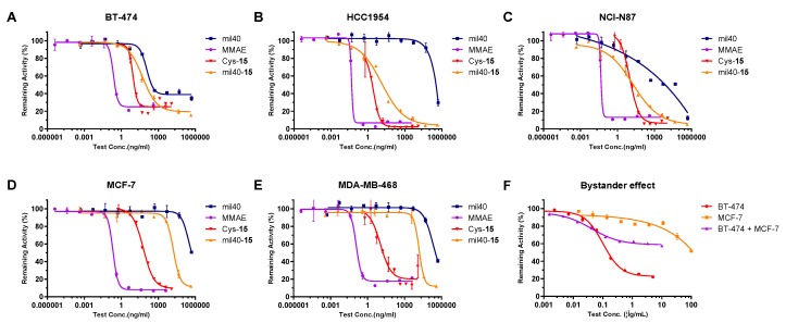
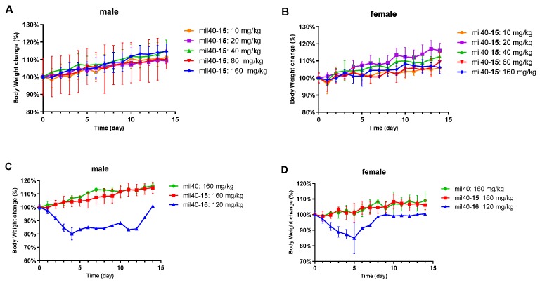
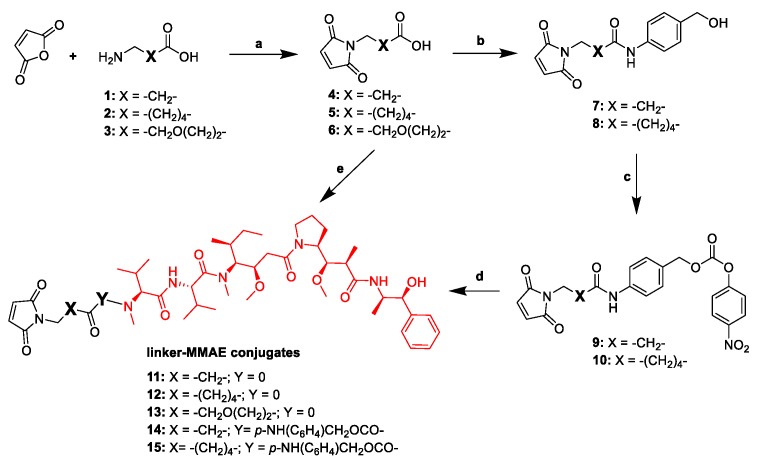
Monomethyl auristatin E (MMAE) is the most popular and widely used cytotoxin in the development of antibody-drug conjugates (ADCs). However, current MMAE-based ADCs are all constructed using cleavable linkers, and this design concept still has insurmountable drawbacks. Their potential instabilities and lipophilic MMAE-induced "bystander effect" inevitably increase the toxicity to normal tissues. Herein, we overturn previous negative views of MMAE-based ADCs with non-cleavable linkers and propose using ionized L-Cysteine (Cys)-linker-MMAE as a novel payload, which can ingeniously enrich and enter tumor cells through receptor-mediated endocytosis of antibodies while its lower permeability helps to avoid further off-target toxicity. We demonstrate that Cys-linker-MMAE maintains high potency similar to free MMAE at the tubulin molecular level and can also be efficiently released in target cells. As a result, the preferred ADC (mil40-15) not only exhibits ideal plasma stability and maintains potent cytotoxicity as MMAE (IC50: 10-11 M), but also shows improved safety with lower bystander toxicity (IC50: 10-9 M), its maximum tolerated dose approaching the level of the naked antibody (160 mg/kg). This study indicated that Cys-linker-MMAE has the potential as a potent payload for ADCs, which is expected to provide novel strategies for the development of MMAE-based ADCs.

Keywords: Cys-linker-MMAE; MMAE; antibody-drug conjugate; linker; tumor.


Conflict of interest statement

The authors declare no conflicts of interest.

Figures

Figure 1
Figure 1
The drug release process of MMAE- and Cys-linker-MMAE-based ADCs. (A) The drug release mechanism of traditional MMAE-based ADCs with cathepsin B-cleavable dipeptide linkers. (B) The drug release mechanism of the novel Cys-linker-MMAE-based ADC and its cellular metabolites. Cys: L-cysteine; MMAE: monomethyl auristatin E; ADCs: antibody-drug conjugates; IC50: half maximal inhibitory concentration; M: mol/L; MlogP: Moriguchi octanol-water partition coefficient.
Figure 2
Figure 2
Tubulin polymerization inhibition analysis. (A) Cys-11, (B) Cys-12, (C) Cys-13, (D) Cys-14, (E) Cys-15 and (F) MMAE were all tested at a range of concentrations (0.1, 0.3, 1.0, 3.0, 10.0, and 30.0 μM), and the inhibitory activity of tubulin polymerization is expressed as the concentration for 50% maximal effect (EC50). The results are shown as the mean ± standard deviation (SD) from triplicate experiments.
Figure 3
Figure 3
Stability of the Cys-linker-MMAE-based ADC in 50% plasma at 37 °C. The test concentration of mil40-15 was 250 nM, and the lower limit of quantification (LLOQ) and upper limit of quantification (ULOQ) of MMAE were 2 nM and 250 nM, respectively. Each sample point contains two repetitions and the results are shown as the mean.
Figure 4
Figure 4
Cytotoxicity of the Cys-linker-MMAE-based ADC in tumor cells. (A) The cytotoxicity assays performed in BT-474 cell line. (B) The cytotoxicity assays performed in HCC1954 cell line. (C) The cytotoxicity assays performed in NCI-N87 cell line. (D) The cytotoxicity assays performed in MCF-7 cell line. (E) The cytotoxicity assays performed in MDA-MB-468 cell line. (F) The bystander effect test performed in BT-474 and MCF-7 cell lines. The results are shown as the mean ± SD from triplicate experiments.
Figure 5
Figure 5
Receptor-mediated internalization of the Cys-linker-MMAE-based ADC in the HER2+ breast cancer cell line BT-474. Cells were treated at 4 °C with mil40 and mil40-15 labeled with an Alexa Fluor 488-conjugated goat anti-human IgG antibody for 0.5 h and further incubated at 37 °C for 16 h. Scale bar = 10 μm.
Figure 6
Figure 6
Xenograft studies of the Cys-linker-MMAE-based ADC. (A) The tumor volume of the test animals during the treatment and observation period. (B) Changes in the body weights of the mice during the observation period. Nude mice were implanted subcutaneously with NCI-N87 cells. When the tumors reached ~120 mm3, the animals were given vehicle, mil40, or ADC on days 0, 7, 14, and 21. The results are shown as the mean ± SD, n = 6/group.
Figure 7
Figure 7
Fluorescence imaging of the Cys-linker-MMAE-based ADC in NCI-N87 xenograft model. (A) In vivo imaging of fluorescein-labeled ADC, color scale represents photons/s/cm2/steradian. (B) The total radiant efficiency of test mice in NCI-N87 xenograft model; the results are shown as the mean ± SD, n = 3/group. (C) The maximum fluorescence of test mice in NCI-N87 xenograft model. (D) In vitro fluorescence imaging of mouse tissues in ADC administration group.
Figure 8
Figure 8
Hematological analysis of the Cys-linker-MMAE-based ADC. The results are shown as the mean ± SD, n = 6/group. WBCs: white blood cells; RBCs: red blood cells; HGB: hemoglobin; HCT: hematocrit; MCV: mean corpuscular volume; MCH: mean corpuscular hemoglobin; PLT: platelets; Neut: neutrophils; Lymph: lymphocytes; Momo: monocytes; Eos: eosinophils; LUC: large unstained cells.
Figure 9
Figure 9
Tolerance study of the Cys-linker-MMAE-based ADC. (A) Body weight changes in male mice after administration of mil40-15. (B) Body weight changes in female mice after administration of mil40-15. (C) Body weight changes in male mice after administration of mil40, mil40-15, and mil40-16. (D) Body weight changes in female mice after administration of mil40, mil40-15, and mil40-16. The results are shown as the mean ± SD, n = 3/group.
Scheme 1
Scheme 1
General synthetic route for the linker-MMAE conjugates. Reagents and conditions: (a) acetic acid, 120 °C, 6–8 h, 74%–86%; (b) p-aminobenzyl alcohol, 1-ethyl-(3-dimethylaminopropyl) carbodiimide hydrochloride (EDCI), N,N′-diisopropylethylamine (DIPEA), dichloromethane (DCM), room temperature (rt), overnight, 33%–55%; (c) bis(4-nitrophenyl) carbonate, DIPEA, N,N′-dimethylformamide (DMF), rt, 5 h, 63%~65%; (d) MMAE, 1-hydroxybenzotriazole (HOBt), DIPEA, DMF, rt, overnight, 50%~90%; (e) MMAE, diethyl cyanophosphonate, DIPEA, DCM, rt, overnight, 55%–63%.

References

    1. Beck A., Goetsch L., Dumontet C., Corvaia N. Strategies and challenges for the next generation of antibody-drug conjugates. Nat. Rev. Drug Discov. 2017;16:315–337. doi: 10.1038/nrd.2016.268. - DOI - PubMed
    1. Johansson M.P., Maaheimo H., Ekholm F.S. New insight on the structural features of the cytotoxic auristatins mmae and mmaf revealed by combined nmr spectroscopy and quantum chemical modelling. Sci. Rep. 2017;7:e15920. doi: 10.1038/s41598-017-15674-1. - DOI - PMC - PubMed
    1. Duerr C., Friess W. Antibody-drug conjugates- stability and formulation. Eur. J. Pharm. Biopharm. 2019;139:168–176. doi: 10.1016/j.ejpb.2019.03.021. - DOI - PubMed
    1. Lyon R. Drawing lessons from the clinical development of antibody-drug conjugates. Drug Discov. Today Technol. 2018;30:105–109. doi: 10.1016/j.ddtec.2018.10.001. - DOI - PubMed
    1. Coats S., Williams M., Kebble B., Dixit R., Tseng L., Yao N.S., Tice D.A., Soria J.C. Antibody-drug conjugates: Future directions in clinical and translational strategies to improve the therapeutic index. Clin. Cancer Res. 2019;25:5441–5448. doi: 10.1158/1078-0432.CCR-19-0272. - DOI - PubMed