. 2020 Mar 21;8(3):447.
doi: 10.3390/microorganisms8030447.

Identification and in vitro Characterization of a Novel Phage Endolysin that Targets Gram-Negative Bacteria

Affiliations

Identification and in vitro Characterization of a Novel Phage Endolysin that Targets Gram-Negative Bacteria

Jaewoo Bai et al. Microorganisms. .

Abstract

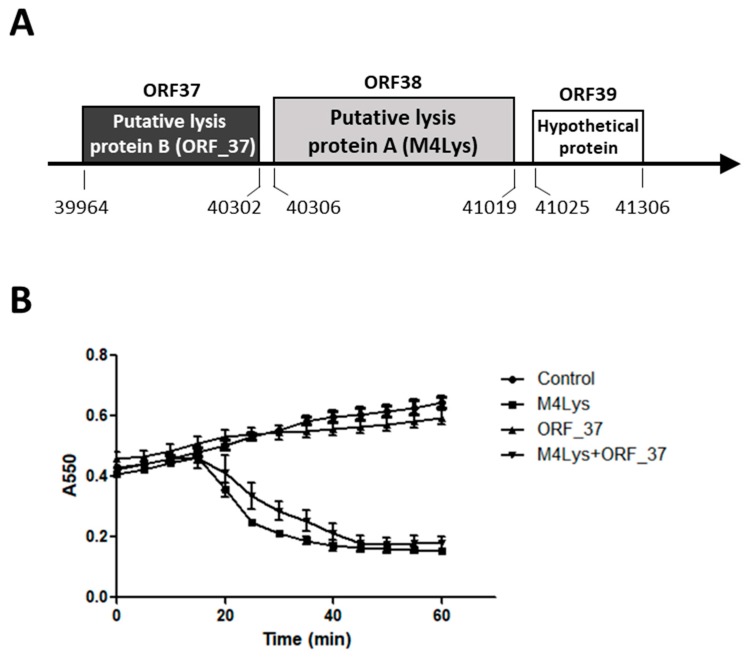
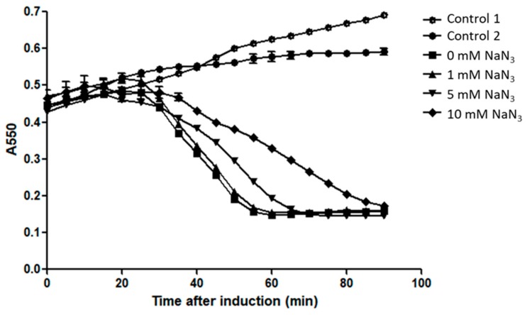
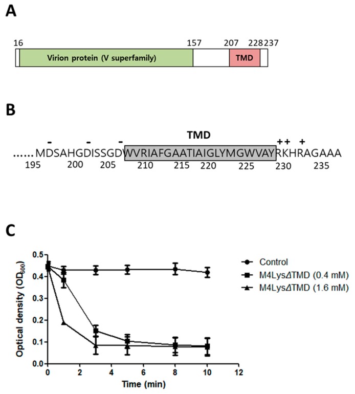
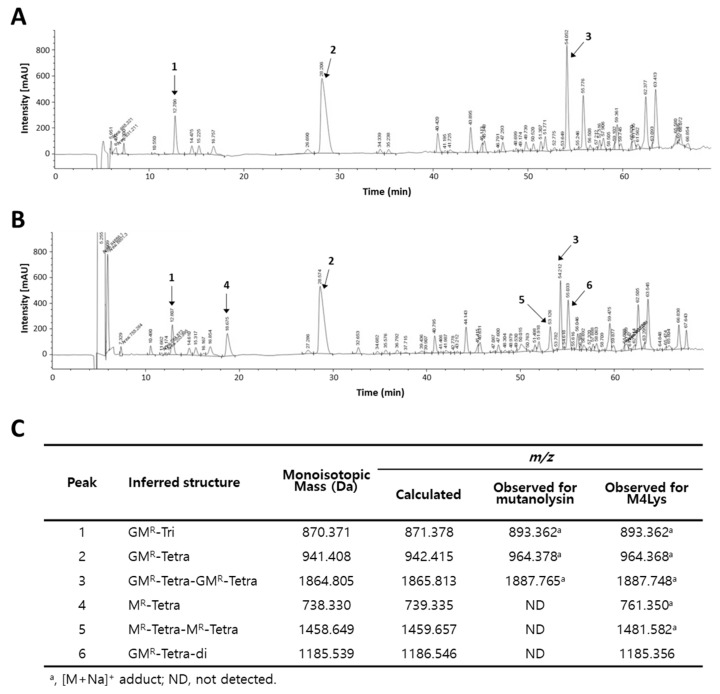
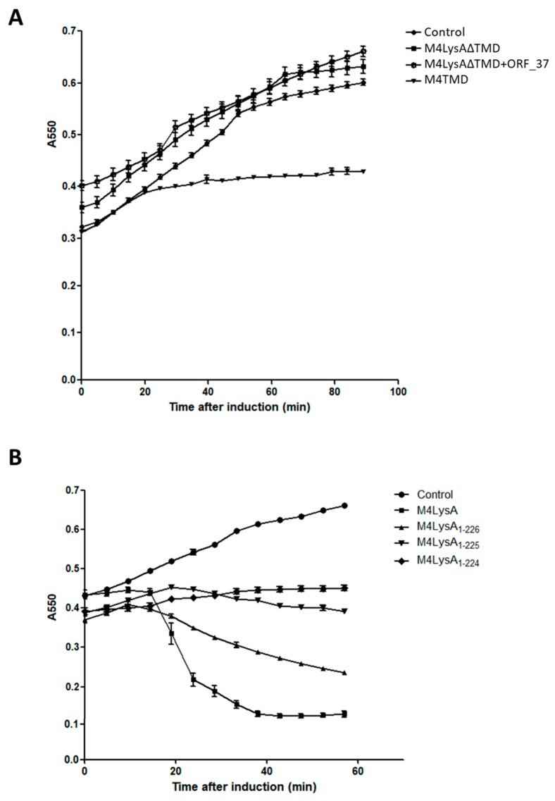
Most double-stranded (ds) DNA phages utilize holin proteins to secrete endolysin for host peptidoglycan lysis. In contrast, several holin-independent endolysins with secretion sequences or signal-arrest-release (SAR) sequences are secreted via the Sec pathway. In this study, we characterized a novel lysis protein (M4Lys) encoded by the dsDNA phage BSPM4, whose lysis function is not dependent on either holin or the Sec pathway in vitro. In silico analysis of M4Lys revealed that it contains a putative virion protein domain and an unusual C-terminal transmembrane domain (TMD). Turbidity reduction assays and liquid chromatography-mass spectrometry using purified peptidoglycan showed that the virion protein domain of M4Lys has peptidoglycan lysis activity. In vitro overproduction of M4Lys in Escherichia coli revealed that M4Lys alone caused rapid cell lysis. Treatment of E. coli with a Sec inhibitor did not inhibit the lysis activity of M4Lys, indicating that the Sec pathway is not involved in M4Lys-mediated cell lysis. Truncation of the TMD eliminated the cell lysis phenomenon, while production of the TMD alone did not induce the cell lysis. All these findings demonstrate that M4Lys is a novel endolysin that has a unique mosaic structure distinct from other canonical endolysins and the TMD plays a critical role in M4Lys-mediated in vitro cell lysis.

Keywords: cell lysis kinetics; endolysin; flagella-targeting phage; secretion; transmembrane domain.


Conflict of interest statement

The authors have no competing financial interests to declare.

Figures

Figure 1
Figure 1
Schematic representation of the phage BSPM4 lysis cassette and analysis of the lysis kinetics by its putative lysis proteins. (A) Schematic representation of the phage BSPM4 lysis cassette; (B) Growth kinetics of the E. coli strains producing the putative lysis protein A (M4Lys) and the putative lysis protein B (ORF_37). Closed circles, empty vector; closed triangles, production of ORF_37; closed squares, production of M4Lys; reverse closed triangles, co-production of ORF_37 and M4Lys. Lysis activity of each gene was investigated by 0.1 mM IPTG induction and the turbidity of the cells was observed. Experiments for the growth kinetics were performed in triplicate. The results of each treatment are represented by the mean ± standard deviation.
Figure 2
Figure 2
Sec machinery is not involved in M4Lys-mediated cell lysis in vitro. Cell lysis kinetics of M4Lys in the presence of the Sec translocase inhibitor, NaN3. In all experiments, expression of M4Lys from pETDuet-1 plasmid in E. coli cells was induced by adding 0.1 mM of IPTG, except for the controls. Open circles, no NaN3 without IPTG (control 1); closed circles, 10 mM NaN3 without IPTG (control 2); closed squares, no NaN3; closed triangles, 1 mM NaN3; reverse triangles, 5 mM NaN3; and diamonds, 10 mM NaN3. All experiments for the cell lysis kinetics were performed in triplicate and the results of each treatment are represented by the mean ± standard deviation.
Figure 3
Figure 3
Domain analysis of M4Lys and its peptidoglycan degradation activity. (A) Domain analysis of M4Lys. The numbers represent amino acid positions; (B) Charge distribution of the TMD region of M4Lys. Positive and negative charges are represented as + and -, respectively, above the amino acid sequences. Numbers under the amino acid sequences indicate amino acid position; (C) Turbidity reduction assay of purified M4LysΔTMD using E. coli peptidoglycan. Closed circles, buffer treatment (control); closed squares, 0.4 mM M4LysΔTMD treatment; and closed triangles, 1.6 mM M4LysΔTMD treatment. Turbidity reduction assays were performed in triplicate and the results of each treatment are represented by the mean ± standard deviation.
Figure 4
Figure 4
Determination of M4Lys cleavage sites. Analysis of E. coli MG1655 peptidoglycan digested by (A) mutanolysin and (B) mutanolysin with purified M4LysΔTMD. Soluble muropeptides were reduced and analyzed by RP-HPLC coupled to MS. Peaks corresponding to m/z values matching previously identified muropeptides are numbered. The fragmentation pattern (peaks 1, 2, and 3) is typical of Tri (L-Ala-D-Glu-m-DAP), Tetra (L-Ala-D-Glu-m-DAP-D-Ala), and Tetra-Tetra muropeptides, respectively. The fragmentation event leading to the loss of a nonreduced GlcNAc residue (203.078, theoretical mass; peaks 4 and 5) indicates the N-acetylmuramidase activity of M4LysΔTMD. Cleavage of D-Glu-mDAP crosslink (peak 6) shows the endopeptidase activity of M4LysΔTMD; (C) Inferred structures, theoretical monoisotopic masses, and theoretical and observed m/z values of individual peaks are tabulated. MR, reduced MurNAc; G, GlcNAc; Di, m-DAP (meso-diaminopimelic acid)-D-Ala; Tri, L-Ala-D-Glu-m-DAP; and Tetra, L-Ala-D-Glu-m-DAP-D-Ala.
Figure 5
Figure 5
Functional analysis of the M4Lys transmembrane domain. (A) Catalytic domain (M4LysΔTMD), C-terminal TMD of M4Lys (M4TMD), and a putative lysis protein B (ORF_37) were produced separately or together in E. coli BL21 (DE3) cells. Closed circles, empty vector; closed square, expression of M4LysΔTMD; open square, coexpression of M4LysΔTMD and ORF_37; and reverse closed triangles, expression of TMD; (B) Lysis activity of various deletion mutants compared with the full-length M4Lys. Closed circles, empty vector; closed squares, full-length M4Lys; closed triangles, 11 amino acid deletion in TMD of M4Lys (M4Lys1–226); reverse closed triangles, 12 amino acid deletion in M4Lys TMD (M4Lys1–225); and closed diamonds, 13 amino acid deletion of M4Lys TMD (M4Lys1–224). Truncation of 13 amino acids from the M4Lys sequence eliminated the lytic activity. All protein expression was induced by adding 0.1 mM IPTG. Experiments for cell lysis kinetics were performed in triplicate and the results of each treatment are represented by the mean ± standard deviation.
Figure 6
Figure 6
Amino acid sequence alignments of M4Lys with other endolysins. (A) Phylogenetic analysis of endolysins from Gram-negative bacteria phages. Each group corresponds to a different functional domain: group I, V superfamily; group II, D-alanyl-D-alanine-carboxypeptidase; group III, N-acetyl muramoyl L-alanine amidase; group IV, glycoside hydrolase family 19; group V, N-acetylmuramidase; and group VI, glycoside hydrolase family 24, respectively; (B) Amino acid sequence alignments of the M4Lys protein of phage BSPM4 with other putative lysis proteins of relevant phages including SPN19, iEPS5, FSLSP088, and Chi. Conserved and identical residues are shaded in gray and black, respectively.
Figure 6
Figure 6
Amino acid sequence alignments of M4Lys with other endolysins. (A) Phylogenetic analysis of endolysins from Gram-negative bacteria phages. Each group corresponds to a different functional domain: group I, V superfamily; group II, D-alanyl-D-alanine-carboxypeptidase; group III, N-acetyl muramoyl L-alanine amidase; group IV, glycoside hydrolase family 19; group V, N-acetylmuramidase; and group VI, glycoside hydrolase family 24, respectively; (B) Amino acid sequence alignments of the M4Lys protein of phage BSPM4 with other putative lysis proteins of relevant phages including SPN19, iEPS5, FSLSP088, and Chi. Conserved and identical residues are shaded in gray and black, respectively.

References

    1. Wang I.N., Smith D.L., Young R. Holins: The protein clocks of bacteriophage infections. Annu. Rev. Microbiol. 2000;54:799–825. doi: 10.1146/annurev.micro.54.1.799. - DOI - PubMed
    1. Bernhardt T.G., Roof W.D., Young R. Genetic evidence that the bacteriophage phi X174 lysis protein inhibits cell wall synthesis. Proc. Natl. Acad. Sci. USA. 2000;97:4297–4302. doi: 10.1073/pnas.97.8.4297. - DOI - PMC - PubMed
    1. Bernhardt T.G., Struck D.K., Young R. The lysis protein E of phi X174 is a specific inhibitor of the MraY-catalyzed step in peptidoglycan synthesis. J. Biol. Chem. 2001;276:6093–6097. doi: 10.1074/jbc.M007638200. - DOI - PubMed
    1. Young I., Wang I., Roof W.D. Phages will out: Strategies of host cell lysis. Trends Microbiol. 2000;8:120–128. doi: 10.1016/S0966-842X(00)01705-4. - DOI - PubMed
    1. Ackermann H.W. Bacteriophage observations and evolution. Res. Microbiol. 2003;154:245–251. doi: 10.1016/S0923-2508(03)00067-6. - DOI - PubMed

LinkOut - more resources