

Invasive cattle ticks in East Africa: morphological and molecular confirmation of the presence of Rhipicephalus microplus in south-eastern Uganda
- PMID: 32245511
- PMCID: PMC7118885
- DOI: 10.1186/s13071-020-04043-z
Invasive cattle ticks in East Africa: morphological and molecular confirmation of the presence of Rhipicephalus microplus in south-eastern Uganda
Abstract
Background: Rhipicephalus microplus, an invasive tick species of Asian origin and the main vector of Babesia species, is considered one of the most widespread ectoparasites of livestock. The tick has spread from its native habitats on translocated livestock to large parts of the tropical world, where it has replaced some of the local populations of Rhipicephalus decoloratus ticks. Although the tick was reported in Uganda 70 years ago, it has not been found in any subsequent surveys. This study was carried out to update the national tick species distribution on livestock in Uganda as a basis for tick and tick-borne disease control, with particular reference to R. microplus.
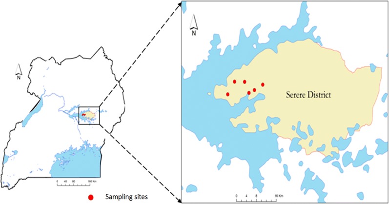
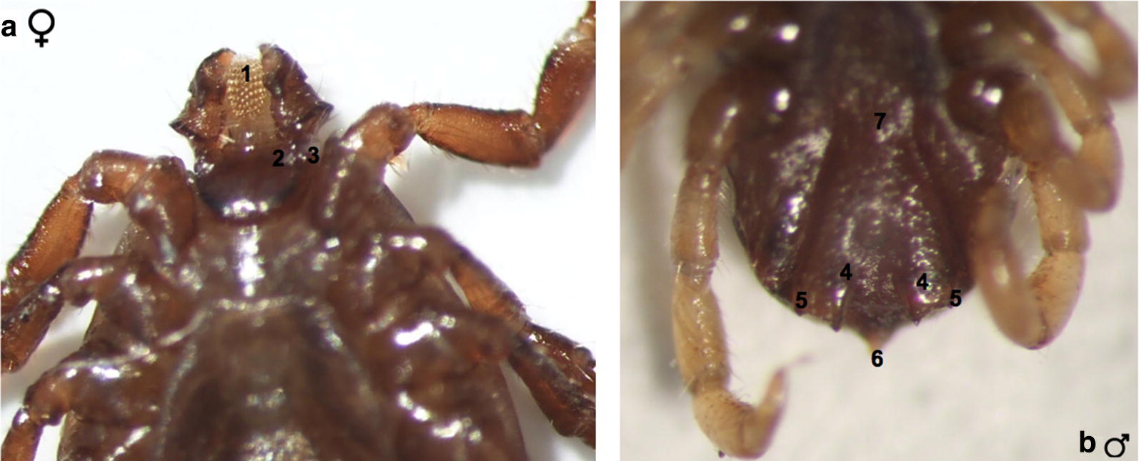
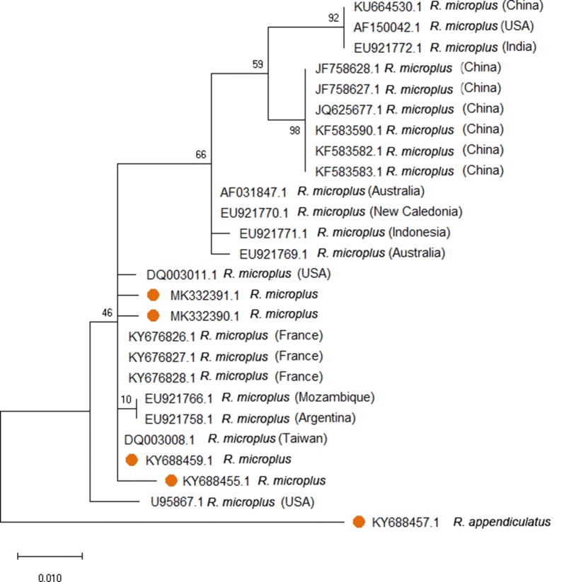
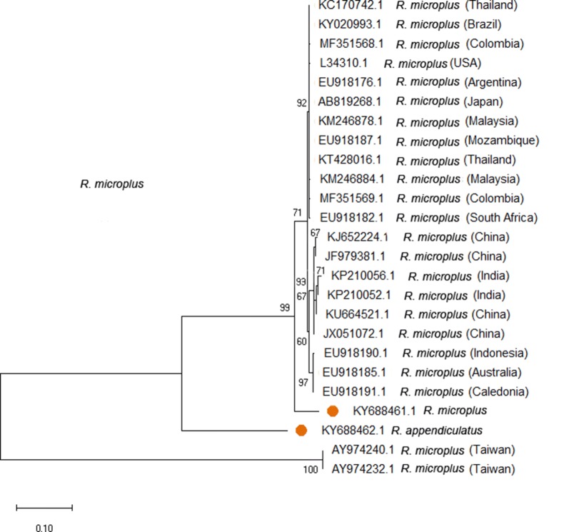
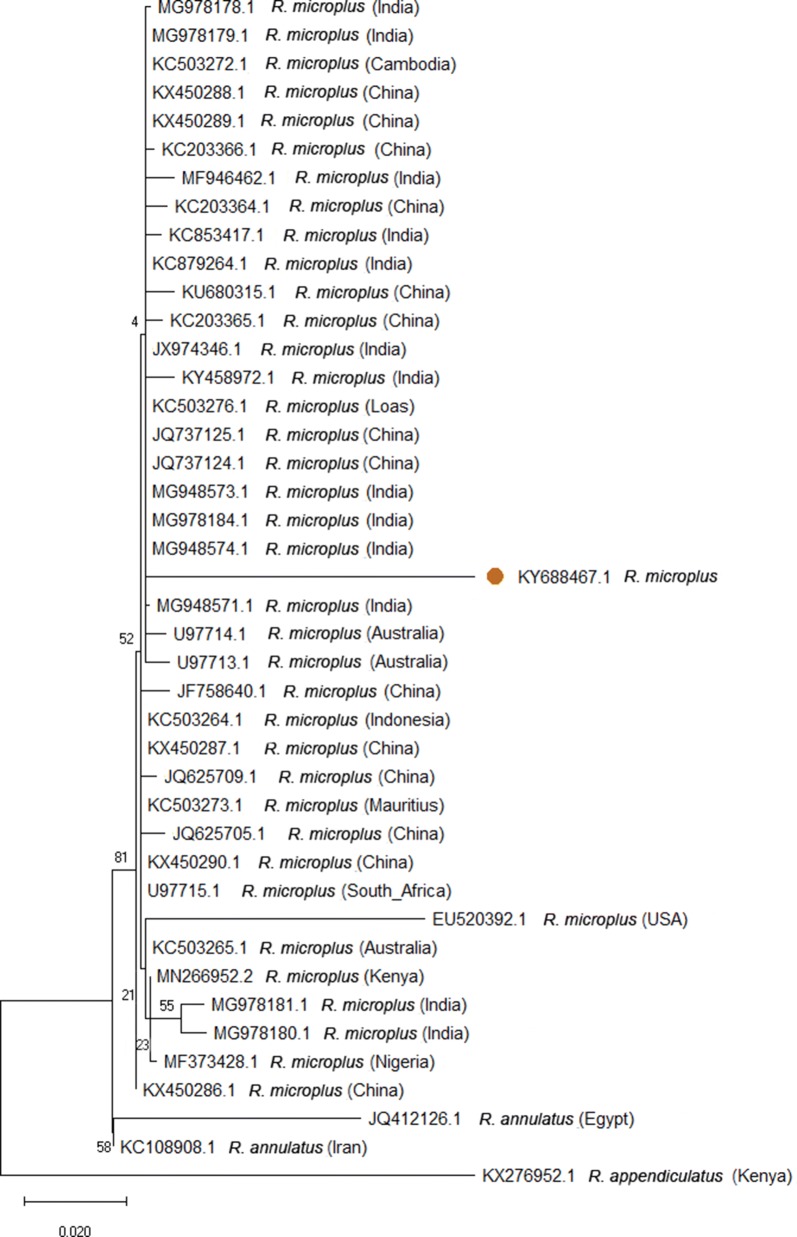
Methods: The study was carried out in Kadungulu, Serere district, south-eastern Uganda, which is dominated by small scale livestock producers. All the ticks collected from 240 cattle from six villages were identified microscopically. Five R. microplus specimens were further processed for phylogenetic analysis and species confirmation.
Results: The predominant tick species found on cattle was Rhipicephalus appendiculatus (86.9 %; n = 16,509). Other species found were Amblyomma variegatum (7.2 %; n = 1377), Rhipicephalus evertsi (2.3 %; n = 434) and R. microplus (3.6 %; n = 687). Phylogenetic analysis of the 12S rRNA, 16S rRNA and ITS2 gene sequences of R. microplus confirmed the morphological identification.
Conclusions: It is concluded that R. microplus has replaced R. decoloratus in the sampled villages in Kadungulu sub-county, since the latter was not any longer found in this area. There is currently no livestock movement policy in force in Uganda, which could possibly limit the further spread of R. microplus ticks. Future surveys, but also retrospective surveys of museum specimens, will reveal the extent of distribution of R. microplus in Uganda and also for how long this tick has been present on livestock without being noticed.
Keywords: Rhipicephalus microplus; Serere district; Tick-borne diseases; Ticks; Uganda.
Conflict of interest statement
The authors declare that they have no competing interests.
Figures
Similar articles
-
Identification and distribution of Rhipicephalus microplus in selected high-cattle density districts in Uganda: signaling future demand for novel tick control approaches.BMC Vet Res. 2024 Mar 25;20(1):119. doi: 10.1186/s12917-024-03979-z. BMC Vet Res. 2024. PMID: 38528496 Free PMC article.
-
Distribution and prevalence of ixodid tick species (Acari: Ixodidae) infesting cattle in Karamoja region of northeastern Uganda.BMC Vet Res. 2024 Feb 7;20(1):50. doi: 10.1186/s12917-023-03802-1. BMC Vet Res. 2024. PMID: 38326882 Free PMC article.
-
A comprehensive survey of the prevalence and spatial distribution of ticks infesting cattle in different agro-ecological zones of Cameroon.Parasit Vectors. 2019 Oct 17;12(1):489. doi: 10.1186/s13071-019-3738-7. Parasit Vectors. 2019. PMID: 31623642 Free PMC article.
-
Review of cattle ticks (Acari, Ixodida) in Ivory Coast and geographic distribution of Rhipicephalus (Boophilus) microplus, an emerging tick in West Africa.Exp Appl Acarol. 2017 Apr;71(4):355-369. doi: 10.1007/s10493-017-0129-7. Epub 2017 May 11. Exp Appl Acarol. 2017. PMID: 28497303 Review.
-
Spread of parasites transported with their hosts: case study of two species of cattle tick.Rev Sci Tech. 2010 Apr;29(1):149-60, 135-47. Rev Sci Tech. 2010. PMID: 20617654 Review. English, French.
Cited by
-
Molecular epidemiology of anaplasmosis in small ruminants along a human-livestock-wildlife interface in Uganda.Heliyon. 2020 Dec 31;7(1):e05688. doi: 10.1016/j.heliyon.2020.e05688. eCollection 2021 Jan. Heliyon. 2020. PMID: 33437885 Free PMC article.
-
Morphological and Molecular Characterization of Tick Species Infesting Cattle in South Africa.Vet Sci. 2024 Dec 10;11(12):638. doi: 10.3390/vetsci11120638. Vet Sci. 2024. PMID: 39728978 Free PMC article.
-
Babesia infection in cattle and dogs in Suizhou City, Hubei Province, China.Infect Med (Beijing). 2025 Feb 21;4(1):100170. doi: 10.1016/j.imj.2025.100170. eCollection 2025 Mar. Infect Med (Beijing). 2025. PMID: 40129442 Free PMC article.
-
Diagnostic performance of a Rapid Tick exposure Test (RaTexT®) to detect acaricide resistance in cattle ticks in East Africa.Parasit Vectors. 2025 Aug 11;18(1):342. doi: 10.1186/s13071-025-06995-6. Parasit Vectors. 2025. PMID: 40790210 Free PMC article.
-
Pathogenic Rickettsia, Anaplasma, and Ehrlichia in Rhipicephalus microplus ticks collected from cattle and laboratory hatched tick larvae.PLoS Negl Trop Dis. 2023 Aug 30;17(8):e0011546. doi: 10.1371/journal.pntd.0011546. eCollection 2023 Aug. PLoS Negl Trop Dis. 2023. PMID: 37647577 Free PMC article.
References
-
- Coetzer JAW, Tustin RC. Infectious diseases of livestock. 2. Cape Town: Oxfor University Press; 2004.
-
- Low VL, Tay ST, Kho KL, Koh FX, Tan TK, Lim YAL, et al. Molecular characterisation of the tick Rhipicephalus microplus in Malaysia: new insights into the cryptic diversity and distinct genetic assemblages throughout the world. Parasit Vectors. 2011;8:341. doi: 10.1186/s13071-015-0956-5. - DOI - PMC - PubMed
MeSH terms
Substances
Grants and funding
LinkOut - more resources
Full Text Sources
