

PI3K/Akt signaling pathway is essential for de novo hair follicle regeneration
- PMID: 32245516
- PMCID: PMC7118821
- DOI: 10.1186/s13287-020-01650-6
PI3K/Akt signaling pathway is essential for de novo hair follicle regeneration
Abstract
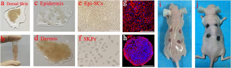
Background: Cultured epidermal stem cells (Epi-SCs) and skin-derived precursors (SKPs) were capable of reconstituting functional hair follicles after implantation, while the signaling pathways that regulate neogenic hair follicle formation are poorly investigated. In this study, we aimed to understand the interactions between Epi-SCs and SKPs during skin organoid formation and to uncover key signal pathways crucial for de novo hair follicle regeneration.
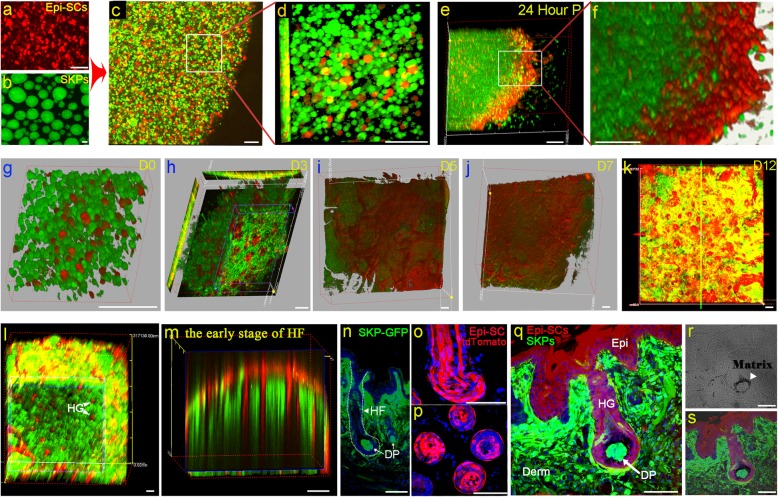
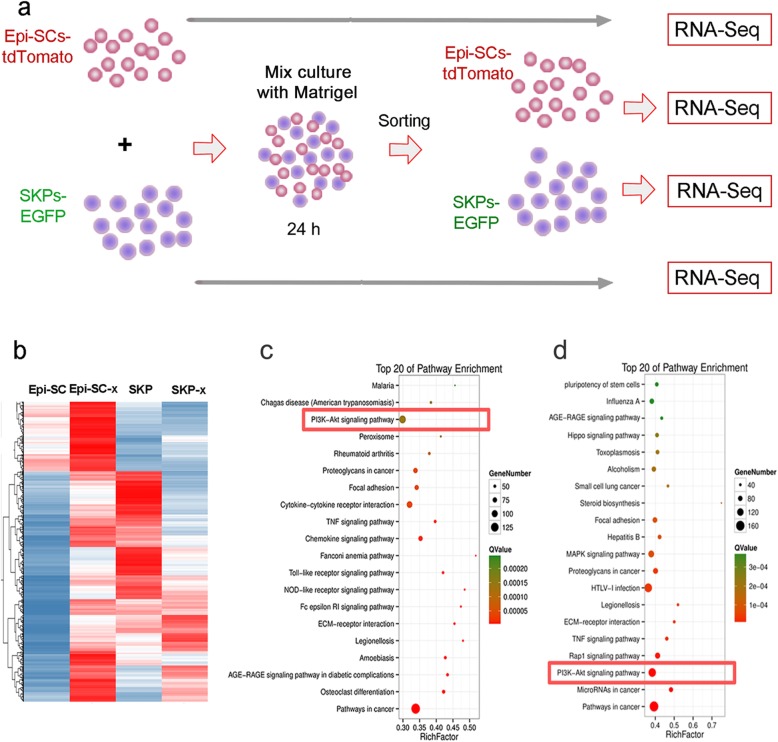
Methods: To track their fate after transplantation, Epi-SCs derived from neonatal C57BL/6 mice were labeled with tdTomato, and SKPs were isolated from neonatal C57BL/6/GFP mice. A mixture of Epi-SCs-tdTomato and SKPs-EGFP in Matrigel was observed under two-photon microscope in culture and after implantation into excisional wounds in nude mice, to observe dynamic migrations of the cells during hair follicle morphogenesis. Signaling communications between the two cell populations were examined by RNA-Seq analysis. Potential signaling pathways revealed by the analysis were validated by targeting the pathways using specific inhibitors to observe a functional loss in de novo hair follicle formation.
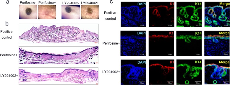
Results: Two-photon microscopy analysis indicated that when Epi-SCs and SKPs were mixed in Matrigel and cultured, they underwent dynamic migrations resulting in the formation of a bilayer skin-like structure (skin organoid), where Epi-SCs positioned themselves in the outer layer; when the mixture of Epi-SCs and SKPs was grafted into excisional wounds in nude mice, a bilayer structure resembling the epidermis and the dermis formed at the 5th day, and de novo hair follicles generated subsequently. RNA-Seq analysis of the two cell types after incubation in mixture revealed dramatic alterations in gene transcriptome, where PI3K-Akt signaling pathway in Epi-SCs was significantly upregulated; meanwhile, elevated expressions of several growth factors and cytokine potentially activating PI3K were found in SKPs, suggesting active reciprocal communications between them. In addition, inhibition of PI3K or Akt by specific inhibitors markedly suppressed the hair follicle regeneration mediated by Epi-SCs and SKPs.
Conclusions: Our data indicate that the PI3K-Akt signaling pathway plays a crucial role in de novo hair follicle regeneration, and the finding may suggest potential therapeutic applications in enhancing hair regeneration.
Keywords: Epi-SCs; Hair follicle regeneration; PI3K-Akt signal; SKPs.
Conflict of interest statement
The authors declare that they have no competing interests.
Figures
Similar articles
-
Hair Follicle and Sebaceous Gland De Novo Regeneration With Cultured Epidermal Stem Cells and Skin-Derived Precursors.Stem Cells Transl Med. 2016 Dec;5(12):1695-1706. doi: 10.5966/sctm.2015-0397. Epub 2016 Jul 25. Stem Cells Transl Med. 2016. PMID: 27458264 Free PMC article.
-
Engineered Skin Substitute Regenerates the Skin with Hair Follicle Formation.Biomedicines. 2021 Apr 8;9(4):400. doi: 10.3390/biomedicines9040400. Biomedicines. 2021. PMID: 33917746 Free PMC article.
-
Amphiregulin promotes hair regeneration of skin-derived precursors via the PI3K and MAPK pathways.Cell Prolif. 2021 Sep;54(9):e13106. doi: 10.1111/cpr.13106. Epub 2021 Aug 12. Cell Prolif. 2021. PMID: 34382262 Free PMC article.
-
Through the lens of hair follicle neogenesis, a new focus on mechanisms of skin regeneration after wounding.Semin Cell Dev Biol. 2020 Apr;100:122-129. doi: 10.1016/j.semcdb.2019.10.002. Epub 2019 Oct 10. Semin Cell Dev Biol. 2020. PMID: 31607627 Free PMC article. Review.
-
Wound-Induced Hair Follicle Neogenesis as a Promising Approach for Hair Regeneration.Mol Cells. 2023 Oct 31;46(10):573-578. doi: 10.14348/molcells.2023.0071. Epub 2023 Aug 31. Mol Cells. 2023. PMID: 37650216 Free PMC article. Review.
Cited by
-
The regulation mechanism of different hair types in inner Mongolia cashmere goat based on PI3K-AKT pathway and FGF21.J Anim Sci. 2022 Nov 1;100(11):skac292. doi: 10.1093/jas/skac292. J Anim Sci. 2022. PMID: 36056739 Free PMC article.
-
Limonin, a Component of Immature Citrus Fruits, Activates Anagen Signaling in Dermal Papilla Cells.Nutrients. 2022 Dec 16;14(24):5358. doi: 10.3390/nu14245358. Nutrients. 2022. PMID: 36558517 Free PMC article.
-
DNA Methylation Dynamics During Esophageal Epithelial Regeneration Following Repair with Acellular Silk Fibroin Grafts in Rat.Adv Biol (Weinh). 2023 May;7(5):e2200160. doi: 10.1002/adbi.202200160. Epub 2023 Jan 19. Adv Biol (Weinh). 2023. PMID: 36658732 Free PMC article.
-
Scoparone Induces Expression of Pluripotency Transcription Factors SOX2 and NANOG in Dermal Papilla Cells.In Vivo. 2021 Sep-Oct;35(5):2589-2597. doi: 10.21873/invivo.12541. In Vivo. 2021. PMID: 34410946 Free PMC article.
-
Pilose antler extracts promotes hair growth in androgenetic alopecia mice by activating hair follicle stem cells via the AKT and Wnt pathways.Front Pharmacol. 2024 Jul 9;15:1410810. doi: 10.3389/fphar.2024.1410810. eCollection 2024. Front Pharmacol. 2024. PMID: 39045053 Free PMC article.
References
-
- Wysocki AB. A review of the skin and its appendages. Adv Wound Care. 1995;8:53–54. - PubMed
Publication types
MeSH terms
Substances
LinkOut - more resources
Full Text Sources
