. 2020 Jun;215(2):421-434.
doi: 10.1534/genetics.120.303052. Epub 2020 Apr 3.

The Dynamics of P Granule Liquid Droplets Are Regulated by the Caenorhabditis elegans Germline RNA Helicase GLH-1 via Its ATP Hydrolysis Cycle

Affiliations

The Dynamics of P Granule Liquid Droplets Are Regulated by the Caenorhabditis elegans Germline RNA Helicase GLH-1 via Its ATP Hydrolysis Cycle

Wenjun Chen et al. Genetics. 2020 Jun.

Abstract

P granules are phase-separated liquid droplets that play important roles in the maintenance of germ cell fate in Caenorhabditis elegans Both the localization and formation of P granules are highly dynamic, but mechanisms that regulate such processes remain poorly understood. Here, we show evidence that the VASA-like germline RNA helicase GLH-1 couples distinct steps of its ATPase hydrolysis cycle to control the formation and disassembly of P granules. In addition, we found that the phenylalanine-glycine-glycine repeats in GLH-1 promote its localization at the perinucleus. Proteomic analyses of the GLH-1 complex with a GLH-1 mutation that interferes with P granule disassembly revealed transient interactions of GLH-1 with several Argonautes and RNA-binding proteins. Finally, we found that defects in recruiting the P granule component PRG-1 to perinuclear foci in the adult germline correlate with the fertility defects observed in various GLH-1 mutants. Together, our results highlight the versatile roles of an RNA helicase in controlling the formation of liquid droplets in space and time.

Keywords: ATP hydrolysis; Germ granule; P granule; RNA helicase; VASA; phase separation.


Figures

Figure 1
Figure 1
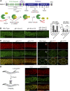
Evidence that germline helicases promote the formation of P granules through their ATP and RNA binding. (A) The domain structure of GLH-1 shows the location of the GLH-1 mutations used in this study (top). The asterisks indicate frameshift deletions that lead to an early stop codon. A model shows how GLH-1 couples its ATP hydrolysis cycle to its RNA binding/release and the proposed effects of the indicated mutations (bottom). (B) GLH-1 and GLH-4 act redundantly to promote the formation of PRG-1 granules. Fluorescent micrographs show the localization of the P granule component GFP::PRG-1 in the indicated strains (left). Image analyses show GFP::PRG-1 granule density and granule size in the indicated strains. Note that uoc1, uoc6, and uoc10 are gene-edited null alleles of the indicated genes. Bar, 10 μm. For the measurements of granule density, the averages and the SDs are indicated. For the measurements of granule size, the median, and 25th and 75th percentiles are indicated. Note that unless specifically labeled, the significance test was measured between wild-type and the indicated strain. *** or ###: P < 0.001. n.s., nonsignificant. (C) The localization of P granule components mCherry::GLH-1 and GFP::PRG-1 is seen in the indicated strains in the adult germline (left) or in the four-cell embryo (right). The arrows indicate the cytoplasmic P granules in the four-cell embryo. Bar, 10 μm. (D) The effects of α-amanitin, an RNA polymerase II inhibitor, on the localization of P granule components mCherry::GLH-1 and GFP::PRG-1 are shown in the adult germline. Images were taken 5 hr after the injection of α-amanitin. Bar, 10 μm.
Figure 2
Figure 2
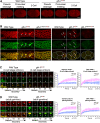
Evidence that germline helicases couple RNA release to promote the turnover of P granule components. (A) The disassembly and the asymmetric sorting of mCherry::GLH-1 granules are defective in early embryos of the glh-1 DQAD mutant. Fluorescent micrographs show the localization of mCherry::GLH-1 at the specified stages of newly fertilized embryos in the indicated strains. Bar, 10 μm. (B) The localization of mCherry::GLH-1 and GFP::PRG-1 in the indicated strains is seen in the adult germline (left) or in the four-cell embryo (right). The arrowheads indicate that many of the enlarged aggregates of P granules in the adult germline do not associate with germline nuclei. The arrows indicate that in embryos, P granules are properly sorted to the cells of the germline lineage (P cell) in wild-type but are improperly sorted to cells of the somatic lineage in the glh-1 DQAD mutant. Bar, 10 μm. (C) Fluorescence recovery after photobleaching (FRAP) analyses indicate that the dynamics of P granule components, including mCherry::GLH-1 and GFP::PRG-1, are both reduced in the four-cell embryos of glh-1 DQAD mutant worms (left). The arrowheads indicate the P granules that are photobleached. Bar, 5 μm. The quantification of FRAP analyses of images showing the average fluorescence signals of mCherry::GLH-1 and GFP::PRG-1 at the indicated times (seconds) after photobleaching (right). Error bars indicated the SDs of fluorescence intensities. (D) FRAP analyses indicate reduced dynamics of P granule components, including GLH-1 and PRG-1, in the adult germlines of glh-1 DQAD mutant worms. Bar, 5 μm. The quantification of FRAP analyses of images shows the average fluorescence signals of mCherry::GLH-1 and GFP::PRG-1 at the indicated times (seconds) after photobleaching (right). Error bars indicate the SD of fluorescence intensities.
Figure 3
Figure 3
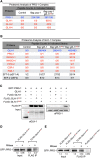
GLH-1 forms a complex that accumulates Argonautes and other RNA-binding proteins in the glh-1 DQAD mutant background. (A) Proteomic analysis of the PRG-1 complex is shown for the indicated strains: wild-type glh-1 and the glh-1 DQAD mutant. The numbers of peptides of the indicated proteins identified in two independent experiments are shown. The proteins whose PRG-1 interactions are stabilized in the glh-1 DQAD mutant are highlighted in red. (B) A proteomic analysis of the GLH-1 complex is given for the indicated glh-1 mutant backgrounds. The proteins whose GLH-1 interactions are stabilized in the glh-1 DQAD mutant are highlighted in red. The numbers of peptides of the indicated proteins identified in two independent experiments are shown. (C) Western blot analyses show the interaction between FLAG::GLH-1 and GFP::PRG-1 is stabilized in the glh-1 DQAD strain. The red asterisk indicates the FLAG::GLH-1. (D) Western blot analyses show that the interaction between FLAG::GLH-1 and GFP::PRG-1 in the glh-1 DQAD mutant is reduced after the RNAse treatment of the purified complex. The number indicates the relative signal intensity. (E) Western blot analyses show that the interaction between FLAG::GLH-1 and GFP::PRG-1 is abolished in the glh-1 DQAD, T663A double mutant and is reduced after the RNAse treatment of the purified complex. The number indicates the relative signal intensity. IP, immunoprecipitation.
Figure 4
Figure 4
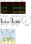
The N-terminal FGG repeats in GLH-1 promote the nuclear anchoring of P granule factors that correlate with fertility. (A) The localization of P granule components GLH-1 and PRG-1 in the adult germline of the indicated strains is seen. Note the strains were grown at their normal growth temperature, 20°. Bar, 10 μm. (B) The brood sizes of worms of the indicated strains are given. Note the strains were grown at an elevated temperature, 25°, which exacerbates their fertility defects (left). The PRG-1 granule density of the indicated strains grown at 20° is shown (middle). The SEM is provided. ***P < 0.001 was determined using one-way ANOVA with the Bonferroni multiple comparison test comparing indicated mutants to wild-type animals. The correlation between brood size and PRG-1 granule density is seen (right). (C) A proposed model shows how GLH-1 controls the dynamics and perinuclear localization of P granules. In this model, the FGG repeats of GLH-1 tether GLH-1 to nuclear pores. GLH-1 then binds newly exported RNAs and facilitates multivalent interactions between the RNAs and RNA-binding proteins to promote P granule formation. As a consequence, GLH-1 couples its RNA binding and release to promote the exchange of P granule factors at the nuclear periphery.

Similar articles

Cited by

References

    1. Alberti S., Saha S., Woodruff J. B., Franzmann T. M., Wang J. et al. , 2018. A user’s guide for phase separation assays with purified proteins. J. Mol. Biol. 430: 4806–4820. 10.1016/j.jmb.2018.06.038 - DOI - PMC - PubMed
    1. Batista P. J., Ruby J. G., Claycomb J. M., Chiang R., Fahlgren N. et al. , 2008. PRG-1 and 21U-RNAs interact to form the piRNA complex required for fertility in C. elegans. Mol. Cell 31: 67–78. 10.1016/j.molcel.2008.06.002 - DOI - PMC - PubMed
    1. Brangwynne C. P., Eckmann C. R., Courson D. S., Rybarska A., Hoege C. et al. , 2009. Germline P granules are liquid droplets that localize by controlled dissolution/condensation. TL - 324. Science 324: 1729–1732. 10.1126/science.1172046 - DOI - PubMed
    1. Claycomb J. M., Batista P. J., Pang K. M., Gu W., Vasale J. J. et al. , 2009. The Argonaute CSR-1 and its 22g-RNA cofactors are required for holocentric chromosome segregation. Cell 139: 123–134. 10.1016/j.cell.2009.09.014 - DOI - PMC - PubMed
    1. Dehghani M., and Lasko P., 2015. In vivo mapping of the functional regions of the DEAD-box helicase Vasa. Biol. Open 4: 450–462. 10.1242/bio.201410579 - DOI - PMC - PubMed

Publication types

MeSH terms

Substances