. 2020 Apr 3;11(1):1658.
doi: 10.1038/s41467-020-15525-0.

Regulatory T cells confer a circadian signature on inflammatory arthritis

Affiliations

Regulatory T cells confer a circadian signature on inflammatory arthritis

L E Hand et al. Nat Commun. .

Abstract

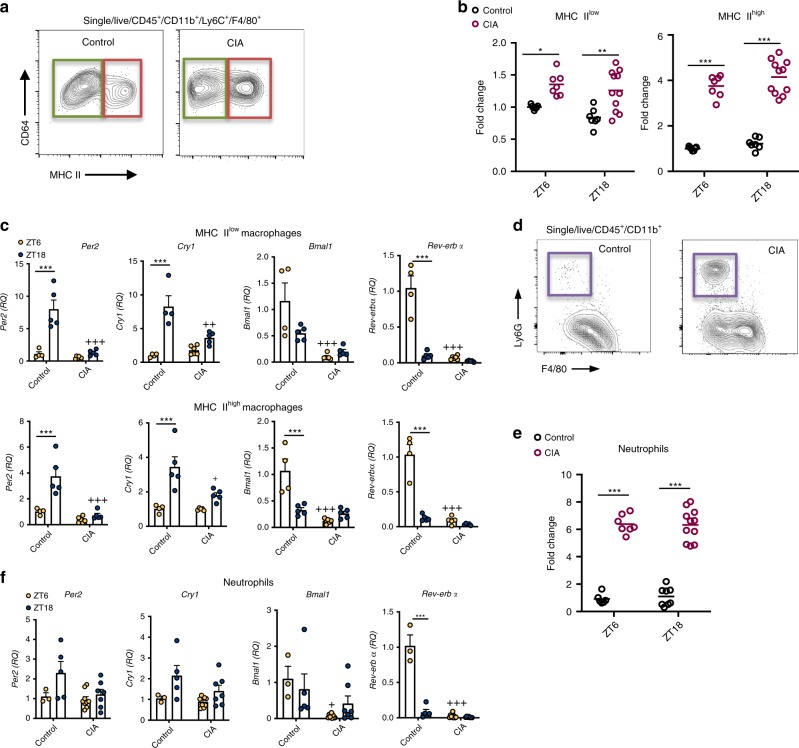
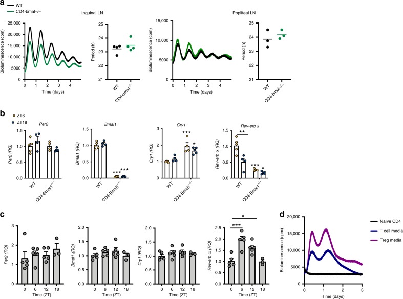
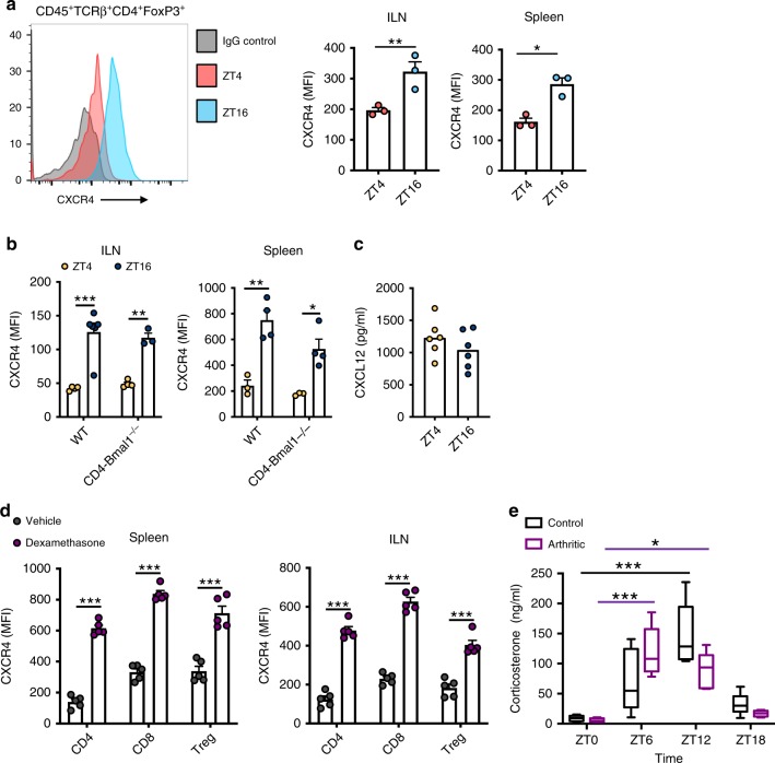
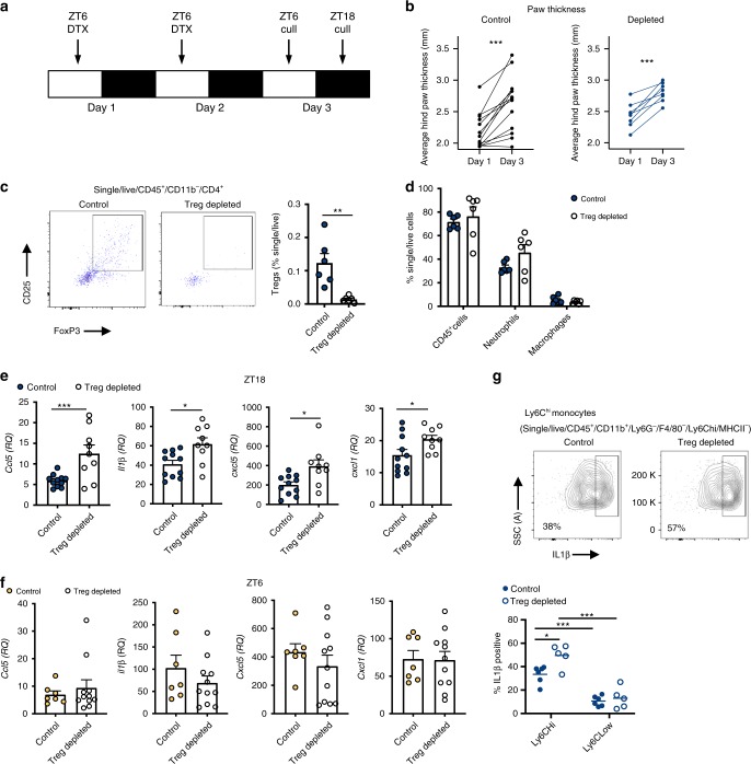
The circadian clock is an intrinsic oscillator that imparts 24 h rhythms on immunity. This clock drives rhythmic repression of inflammatory arthritis during the night in mice, but mechanisms underlying this effect are not clear. Here we show that the amplitude of intrinsic oscillators within macrophages and neutrophils is limited by the chronic inflammatory environment, suggesting that rhythms in inflammatory mediators might not be a direct consequence of intrinsic clocks. Anti-inflammatory regulatory T (Treg) cells within the joints show diurnal variation, with numbers peaking during the nadir of inflammation. Furthermore, the anti-inflammatory action of Treg cells on innate immune cells contributes to the night-time repression of inflammation. Treg cells do not seem to have intrinsic circadian oscillators, suggesting that rhythmic function might be a consequence of external signals. These data support a model in which non-rhythmic Treg cells are driven to rhythmic activity by systemic signals to confer a circadian signature to chronic arthritis.


Conflict of interest statement

The authors declare no competing interests.

Figures

Fig. 1
Fig. 1. Macrophage and neutrophil rhythms under chronic inflammation.
a Two distinct macrophage populations were identified within joints, MHC IIlow (green box) and MHC IIhigh (red box). b Numbers of MHC IIlow and MHCIIhigh macrophages increased within the joints of arthritic animals, neither showed any time-of-day variation in numbers under control or arthritic conditions, data pooled from two separate experiments and normalised to control ZT6 mice (ZT6: control n = 7; CIA n = 7; ZT18: control n = 7; CIA n = 11), two-way ANOVA and Bonferonni post hoc tests. Graphs show individual data points with mean values. c MHC IIlow and MHC IIhigh macrophages sorted from joints at ZT6 (control n = 4; CIA n = 6) or ZT18 (control n = 5; CIA n = 5) were analysed for clock gene expression (normalised to gapdh), data normalised to control ZT6, two-way ANOVA and post-hoc Bonferonni. Data are presented as mean values ± SEM. d Representative flow cytometry plots showing abundant neutrophil infiltration into the joints in CIA. e Numbers of neutrophils increased within the joints of arthritic animals, but did not show time-of-day variation in numbers under control or arthritic conditions, data pooled from two separate experiments and normalised to control ZT6 mice (ZT6: control n = 6; CIA n = 9; ZT18: control n = 8; CIA n = 11), two-way ANOVA and Bonferonni post hoc tests. Graph shows individual data points with mean values. f Clock gene expression (normalised to gapdh) in neutrophils sorted from joints at ZT6 (control n = 3; CIA n = 9) and ZT18 (control n = 5; CIA n = 7), data normalised to control ZT6, two-way ANOVA and post-hoc Bonferonni. Data are presented as mean values ± SEM. In all panels statistical significance between timepoints is shown as *p < 0.05, **p < 0.01 and ***p < 0.005 and significant statistical differences between treatment at given timepoints as +p < 0.05, ++p < 0.01 and +++p < 0.005. Source data are provided as a Source Data file.
Fig. 2
Fig. 2. Diurnal variation in T cell populations within inflamed joints.
a CD3ε T cells (CD45+/CD11b/CD3ε+) in limbs harvested from control and arthritic mice at ZT6 (control n = 7; CIA n = 5) and ZT18 (control n = 7; CIA, n = 8), data normalised to CD3ε T cell numbers in control limbs at ZT6, two-way ANOVA and Bonferonni post hoc tests. b Quantification of CD4+ T cell subsets (Th1 and Th17) in limbs at ZT6 (Th1: control n = 7; CIA n = 13; Th17: control n = 8; CIA n = 13) and ZT18 (Th1: control n = 5; CIA n = 12; Th17: control n = 8; CIA n = 12), data pooled from two separate experiments and normalised to ZT6 control animals, two-way ANOVA and post hoc Bonferonni. c Quantification of Tregs in limbs at ZT6 (control n = 8; CIA n = 13) and ZT18 (control n = 8; CIA n = 12), data pooled from two separate experiments and normalised to ZT6 control animals, two-way ANOVA and post hoc Bonferonni. d Expression of the proliferation markers Ki67 (ZT6: control n = 4; CIA n = 7; ZT18: control n = 4; CIA n = 13) and EdU (ZT6: control n = 6; CIA n = 8; ZT18: control n = 10; CIA n = 8) on Tregs isolated from control and inflamed joints, Kruskal–Wallis test and post-hoc Dunn’s test. e Expression of NRP1 and GITR (ZT6: control n = 4; CIA n = 6; ZT18: control n = 4; CIA n = 12) on Tregs isolated from control and inflamed joints, two-way ANOVA and post-hoc Bonferonni test. NRP1—interaction between time-of-day and disease effect (p = 0.0285); GITR—disease effect (p = 0.0077). f IL10 expression by stimulated Tregs isolated from inflamed joints at ZT6 (n = 8) and ZT18 (n = 17) expressed as total number of IL10+ Tregs and % of Tregs that are IL10+, Mann–Whitney test. g Quantification of Tregs in blood of arthritic mice (ZT6 n = 3; ZT18 n = 6), two-sided unpaired T-test. h Quantification of Tregs in inguinal (ZT6 n = 7; ZT18 n = 5) and popliteal (ZT6 n = 5; ZT18 n = 4) lymph nodes from arthritic mice. Treg numbers were determined per lymph node, only popliteal lymph nodes draining inflamed limbs were analysed, two-sided unpaired T tests. All graphs show individual data points with mean values. In all panels statistical significance between indicated groups is shown as *p < 0.05, **p < 0.01 and ***p < 0.005. Source data are provided as a Source Data file.
Fig. 3
Fig. 3. Treg cells do not have an intrinsic clock.
a Representative PMT traces and calculated period from paired inguinal (n = 4) and paired popliteal (n = 3) lymph nodes from CD4-Bmal1−/− and wildtype mice on a PER2::luc background. Graphs show individual data points and mean values. b Tregs were sorted from the spleens of wildtype and CD4-Bmal1−/− mice at ZT6 (WT n = 5; CD4-Bmal1−/− n = 4) and ZT18 (WT n = 4; CD4-Bmal1−/− n = 5) and utilised for QPCR to assess clock gene expression (normalised to gapdh), two-way ANOVA and post hoc Bonferonni. Unless indicated otherwise, significance shown compares genotype by time-of-day. Data are presented as mean values ± SEM. c Tregs were sorted from lymph nodes of mice at four time points across the day, identified by CD25+CD127low. Expression of clock genes (normalised to gapdh) was quantified by QPCR, normalised to ZT0 (ZT0,6,12 n = 5; ZT18 n = 3), one-way ANOVA and post hoc Bonferonni. Data are presented as mean values ± SEM. d PMT traces of naïve CD4+ T cells purified from the lymph nodes of PER2::luc mice and cultured with IL2 alone, T cell media or Treg media, representative of three independent repeats. In all panels statistical significance is shown as *p < 0.05, **p < 0.01 and ***p < 0.005. Source data are provided as a Source Data file.
Fig. 4
Fig. 4. Glucocorticoid action on Treg cells.
a CXCR4 expression (geometric mean fluorescent intensity) on Tregs from spleen and inguinal lymph nodes of wildtype mice harvested at ZT4 (n = 3) or ZT16 (n = 3) two-sided unpaired T-test. Data are presented as mean values ± SEM. b Diurnal rhythmicity in CXCR4 expression persists in the absence of Bmal1 ILN (ZT4: WT n = 4; CD4-Bmal1−/− n = 4; ZT16: WT n = 6; CD4-Bmal1−/− n = 3) and spleen (ZT4: WT n = 3; CD4-Bmal1−/− n = 3; ZT16: WT n = 4; CD4-Bmal1−/− n = 4), two-way ANOVA, post hoc Tukey. Data are presented as mean values ± SEM. c Plasma levels of CXCL12 in naïve mice at ZT4 and ZT16 (n = 6/group). Graph shows individual data points and mean values. d Effects of 4 h in vivo application of dexamethasone (2 mg/kg) or vehicle (cyclodextrin) at ZT0 on CXCR4 expression on the surface of T cell subsets derived from spleen or inguinal lymph nodes (n = 5/group). One-way ANOVA and post hoc Bonferonni. Data are presented as mean values ± SEM. e Plasma corticosterone levels in naïve (n = 6/time point) and arthritic (ZT0 n = 4; ZT6 n = 5; ZT12 n = 5; ZT18 n = 4) animals across a 24 h period (centre of box represents the medium and bounds extend from 25th to 75th percentile, whiskers are minima and maxima), two-way ANOVA (interaction between time-of-day and disease effect p = 0.0058; Time-of-day effect p < 0.0001; Disease effect NS) and post-hoc Tukey. In panels ad statistical significance is shown as *p < 0.05, **p < 0.01 and ***p < 0.005. In panel e *p < 0.05 and ***p < 0.005 indicates significant statistical differences between the indicated time-points in naïve animals (black lines) and arthritic animals (purple lines). Source data are provided as a Source Data file.
Fig. 5
Fig. 5. Depletion of Treg cells increases localised proinflammatory cytokine expression.
a Two daily injections of DTX at ZT6 led to significant depletion of Tregs 24–36 h after the last injection. b Progression of arthritis (as assessed by hind paw swelling) in control mice (n = 13) and in mice with depleted Tregs (n = 8), two-sided paired T tests. c Quantification of Tregs in arthritic control (n = 6) and Treg depleted (n = 6) animals, two-sided unpaired T-tests. Data are presented as mean values ± SEM. d Quantification of inflammatory cell populations within the inflamed joints in arthritic control (n = 6) and Treg-depleted mice (n = 6), one-way ANOVA. Data are presented as mean values ± SEM. e QPCR analysis of inflammatory genes (normalised to gapdh) within the joints of arthritic control (n = 11) and Treg-depleted (n = 9) mice culled at ZT18. Values were normalised to expression in control animals without arthritis, two-sided unpaired t tests. Data are presented as mean values ± SEM. f QPCR analysis of inflammatory genes (normalised to gapdh) within the joints of arthritic control (n = 7) and Treg-depleted (n = 11) mice culled at ZT6. Values were normalised to expression in control animals without arthritis. Data are presented as mean values ± SEM. g Effect of depletion of Tregs on IL-1β expression by joint monocytes (control n = 6; Treg depleted n = 5), two-way ANOVA and post hoc Bonferonni. Graph shows individual data points and mean values. In all panels statistical significance is shown as *p < 0.05, **p < 0.01 and ***p < 0.005. Source data are provided as a Source Data file.

References

    1. Scheiermann C, Gibbs J, Ince L, Loudon A. Clocking in to immunity. Nat. Rev. Immunol. 2018;18:423–437. doi: 10.1038/s41577-018-0008-4. - DOI - PubMed
    1. Druzd D, et al. Lymphocyte circadian clocks control lymph node trafficking and adaptive immune responses. Immunity. 2017;46:120–132. doi: 10.1016/j.immuni.2016.12.011. - DOI - PMC - PubMed
    1. Suzuki K, Hayano Y, Nakai A, Furuta F, Noda M. Adrenergic control of the adaptive immune response by diurnal lymphocyte recirculation through lymph nodes. J. Exp. Med. 2016;213:2567–2574. doi: 10.1084/jem.20160723. - DOI - PMC - PubMed
    1. Nguyen KD, et al. Circadian gene Bmal1 regulates diurnal oscillations of Ly6C(hi) inflammatory monocytes. Science. 2013;341:1483–1488. doi: 10.1126/science.1240636. - DOI - PMC - PubMed
    1. Keller M, et al. A circadian clock in macrophages controls inflammatory immune responses. Proc. Natl Acad. Sci. USA. 2009;106:21407–21412. doi: 10.1073/pnas.0906361106. - DOI - PMC - PubMed

Publication types