. 2020 Apr 3;11(1):1656.
doi: 10.1038/s41467-020-15517-0.

Mechanism of ribosome shutdown by RsfS in Staphylococcus aureus revealed by integrative structural biology approach

Affiliations

Mechanism of ribosome shutdown by RsfS in Staphylococcus aureus revealed by integrative structural biology approach

Iskander Khusainov et al. Nat Commun. .

Abstract

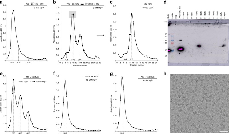
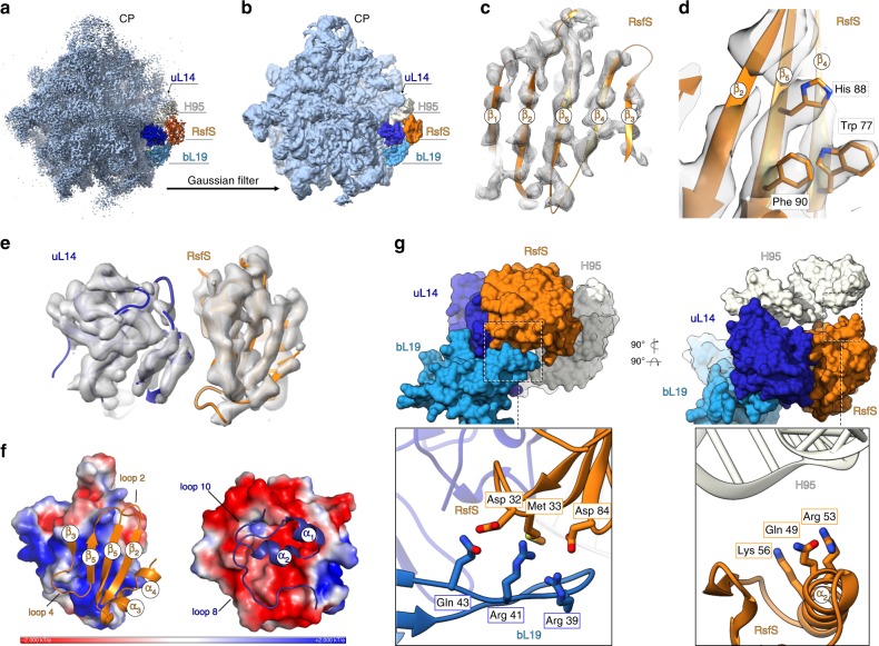
For the sake of energy preservation, bacteria, upon transition to stationary phase, tone down their protein synthesis. This process is favored by the reversible binding of small stress-induced proteins to the ribosome to prevent unnecessary translation. One example is the conserved bacterial ribosome silencing factor (RsfS) that binds to uL14 protein onto the large ribosomal subunit and prevents its association with the small subunit. Here we describe the binding mode of Staphylococcus aureus RsfS to the large ribosomal subunit and present a 3.2 Å resolution cryo-EM reconstruction of the 50S-RsfS complex together with the crystal structure of uL14-RsfS complex solved at 2.3 Å resolution. The understanding of the detailed landscape of RsfS-uL14 interactions within the ribosome shed light on the mechanism of ribosome shutdown in the human pathogen S. aureus and might deliver a novel target for pharmacological drug development and treatment of bacterial infections.


Conflict of interest statement

The authors declare no competing interests.

Figures

Fig. 1
Fig. 1. RsfS protein binds to the 50S particles and prevents ribosomal subunits association.
a Sucrose gradient (SG) profile of the 70S at semi-dissociation conditions (3 mM Mg2+) with the equilibrium shifted toward the association. Bold marker indicates the fraction “70S” taken for the western blot analysis. b, c Purification of the 50S–RsfS complex for cryo-EM studies. SG profile of the 70S at 3 mM Mg2+ upon addition of 5X excess of RsfS (b), and subsequent centrifugation of the complex at 10 mM Mg2+ (c). The gray square indicates the fractions pooled for purification. Bold markers indicate the fractions taken for the western blot analysis. The peak of the 50S–RsfS complex (c) was taken for cryo-EM grid preparation and MS analysis. d Western blot analysis of the selected fractions. e SG profile of 70S + RsfS mixture that was incubated at 3 mM and spun at 10 mM Mg2+. fg SG profiles of the 70S mixed 5× (f) or 15× (g) molar excess of RsfS, incubated and spun at 10 mM Mg2+. h Typical electron micrograph of the 50S–RsfS sample. Scale bar represents 50 nm. Source data for panels ac and eg are provided as a Source Data file.
Fig. 2
Fig. 2. Cryo-EM reconstruction of the 50S–RsfS complex and model interpretation.
a The 3.2 Å cryo-EM density map. Ribosomal protein uL14 is colored in dark blue, bL19 in bright blue, 23S rRNA Helix 95 in white, and RsfS in orange. CP central protuberance. b A low-pass filtered map of the 50S–RsfS complex (Gaussian filter with the width equal to 3.5 voxel size of the initial map) was used for the initial flexible fitting of the molecular model. c, d Density corresponding to the RsfS beta-sheet assembly and model is fitting. For representation reasons, a Gaussian filter with the width equal to one voxel size (outer mesh) was applied to the initial cryo-EM map (inner mesh). e The uL14–RsfS interaction interface as seen in the cryo-EM map (a Gaussian filter with the width equal to 1.5 voxel size applied). f Electrostatic potential of uL14 and RsfS (surface representation) calculated from the model; the structural elements involved in contacts formation are shown as ribbons and labeled. g The RsfS-binding cavity, including uL14, bL19, and H95. Molecules are shown in surface representation. Close-up sections demonstrate potential interacting residues of the proteins (represented as sticks).
Fig. 3
Fig. 3. The crystal structure of uL14–RsfS complex and their interaction interface.
a Atomic coordinates of one of the two uL14–RsfS heterodimers from the asymmetric unit. The Cα root-mean-square deviation (RMSD) between the two heterodimers was calculated to be 0.585 Å, suggesting that these two copies are essentially identical. The uL14 is shown in cyan, RsfS in gold; N and C stand for N-terminus and C-terminus of the proteins. b Amino acids of uL14 and RsfS proteins involved in contact formation. Hydrophobic regions are shown as yellow surfaces, contacting amino acids as sticks. Yellow, red, and blue frames show the H-bonds-forming amino acids form the respective regions marked on surface representation. H-bonds are shown as black dashed lines; the water molecules are shown as red ball; the crystallographic electron density is shown in mesh at 1.5 sigma value.
Fig. 4
Fig. 4. Eukaryotic initiation factor eIF6 shares a similar binding site on universal ribosomal protein uL14, but has a different structure.
a Localization of RsfS and eIF6 proteins on S. aureus 50S and Tetrahymena thermophila 60S, respectively. For a visual representation, the maps were simulated from the atomic coordinates of the models. Ribosomal RNAs are colored in light blue, ribosomal proteins in blue, S. aureus uL14 in dark blue, RsfS in orange, T. thermophila uL14 in yellow, and eIF6 in green. Individual proteins are shown in ribbon to represent their interaction interface, which displays the structural similarity of uL14 and the dissimilarity of the RsfS and eIF6 proteins in the two organisms. Residues of uL14 involved in the interaction with RsfS/eIF6 are shown as sticks, and labeled accordingly. b Pairwise sequence alignment of uL14 from S. aureus and T. thermophila demonstrates their high sequence similarity. Secondary structure elements are shown above the alignment. Residues interacting with RsfS (or eIF6) are highlighted in bold. The RMSD (given in Å) of 3D structure alignment is shown as bars above the sequence. An asterisk indicates a single, fully conserved residue (identity), colon indicates conservation between amino acids with substantial chemical similarity, while dot indicates conservation between amino acids with poor chemical similarity.

References

    1. Starosta AL, Lassak J, Jung K, Wilson DN. The bacterial translation stress response. FEMS Microbiol. Rev. 2014;38:1172–1201. doi: 10.1111/1574-6976.12083. - DOI - PMC - PubMed
    1. Prossliner T, Skovbo Winther K, Sørensen MA, Gerdes K. Ribosome hibernation. Annu. Rev. Genet. 2018;52:321–348. doi: 10.1146/annurev-genet-120215-035130. - DOI - PubMed
    1. Rae CD, Gordiyenko Y, Ramakrishnan V. How a circularized tmRNA moves through the ribosome. Science. 2019;363:740–744. doi: 10.1126/science.aav9370. - DOI - PMC - PubMed
    1. Neubauer C, Gillet R, Kelley AC, Ramakrishnan V. Decoding in the absence of a codon by tmRNA and SmpB in the ribosome. Science. 2012;335:1366–1369. doi: 10.1126/science.1217039. - DOI - PMC - PubMed
    1. Gagnon MG, Seetharaman SV, Bulkley D, Steitz TA. Structural basis for the rescue of stalled ribosomes: structure of YaeJ bound to the ribosome. Science. 2012;335:1370–1372. doi: 10.1126/science.1217443. - DOI - PMC - PubMed

Publication types