. 2020 Apr 3;10(1):5889.
doi: 10.1038/s41598-020-62039-2.

Targeting of radioactive platinum-bisphosphonate anticancer drugs to bone of high metabolic activity

Affiliations

Targeting of radioactive platinum-bisphosphonate anticancer drugs to bone of high metabolic activity

Robin A Nadar et al. Sci Rep. .

Abstract

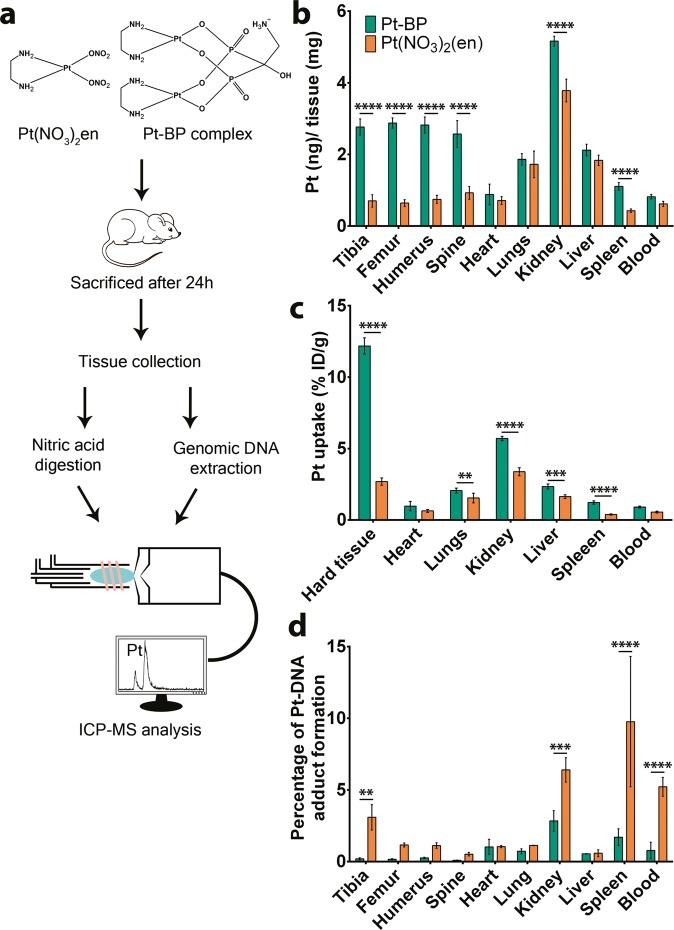
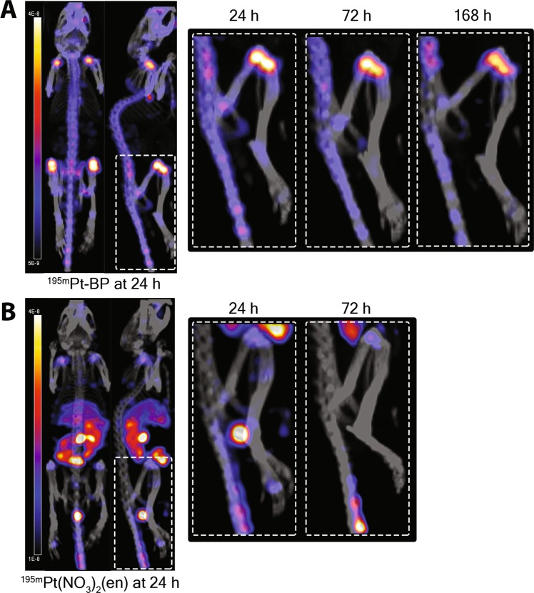
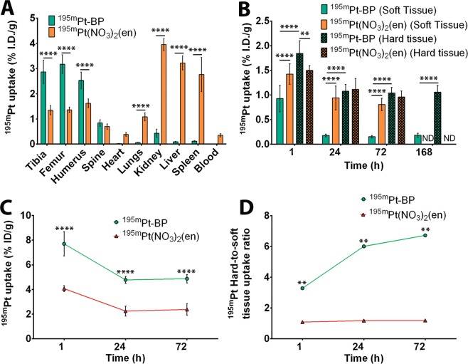
Platinum-based chemotherapeutics exhibit excellent antitumor properties. However, these drugs cause severe side effects including toxicity, drug resistance, and lack of tumor selectivity. Tumor-targeted drug delivery has demonstrated great potential to overcome these drawbacks. Herein, we aimed to design radioactive bisphosphonate-functionalized platinum (195mPt-BP) complexes to confirm preferential accumulation of these Pt-based drugs in metabolically active bone. In vitro NMR studies revealed that release of Pt from Pt BP complexes increased with decreasing pH. Upon systemic administration to mice, Pt-BP exhibited a 4.5-fold higher affinity to bone compared to platinum complexes lacking the bone-seeking bisphosphonate moiety. These Pt-BP complexes formed less Pt-DNA adducts compared to bisphosphonate-free platinum complexes, indicating that in vivo release of Pt from Pt-BP complexes proceeded relatively slow. Subsequently, radioactive 195mPt-BP complexes were synthesized using 195mPt(NO3)2(en) as precursor and injected intravenously into mice. Specific accumulation of 195mPt-BP was observed at skeletal sites with high metabolic activity using micro-SPECT/CT imaging. Furthermore, laser ablation-ICP-MS imaging of proximal tibia sections confirmed that 195mPt BP co-localized with calcium in the trabeculae of mice tibia.


Conflict of interest statement

The authors declare no competing interests.

Figures

Figure 1
Figure 1
Biodistribution profile of Pt-BP and Pt(NO3)2(en) in vivo. (A) Schematic representation of Pt biodistribution study in C57Bl/6N mice. Pt-BP or Pt(NO3)2(en) compounds were administered intravenously in mice (2.5 mM Pt concentration) followed by sacrifice after 24 h. Hard tissues (tibia, femur, humerus, spine), soft tissues (heart, lungs, kidney, liver, spleen) and blood were collected. Collected tissues were subjected to nitric acid digestion and genomic DNA extraction for Pt quantification using ICP-MS and high-resolution ICP-MS, respectively. (B) Pt concentration in different tissues was normalized for specific tissue weight 24 h after injection. Data from 5 mice per group are represented as ng Pt/mg tissue ± SD. (C) Percentage of Pt uptake in different tissues 24 h after injection. Data from 5 mice per group are represented as % I.D./g ± SD of specific tissue type. (D) Percentage of Pt-DNA adduct formation relative to total Pt uptake in specific tissue as quantified by High Resolution-ICP-MS. Data from 4–5 mice per group are presented. **P < 0.01; ***P < 0.001; ****P < 0.0001 as determined by two-way ANOVA with a Bonferroni (multiple comparisons) post-hoc test.
Figure 2
Figure 2
Biodistribution profile of 195mPt-BP and 195mPt(NO3)2(en) in vivo. Representative whole-body micro-SPECT/CT images of biodistribution of 195mPt-BP (A) and 195mPt(NO3)2(en) (B) in nude mice 24 hours after systemic administration. Rectangles show specific biodistribution of these compounds in long bones (femur-tibia) 24 h, 72 h and 168 h after systemic administration.
Figure 3
Figure 3
Quantification of 195mPt-BP and 195mPt(NO3)2(en) biodistribution in vivo. (A) Percentage of injected dose (%ID/g) of 195mPt-BP (after 7 days) and 195mPt(NO3)2(en) (after 3 days) in mice determined using gamma counting. (B) Percentage of injected dose (%ID/g) of 195mPt-BP and 195mPt(NO3)2(en) in mice as quantified from the micro-SPECT/CT images in soft and hard tissues. (C) Percentage of injected dose (%ID/g) of 195mPt-BP and 195mPt(NO3)2(en) in mice as quantified from the micro-SPECT/CT images in the hard tissue region of interest (ROI) with the location of the edge of the ROI contour representing 75% of maximum intensity. (D) 195mPt hard-to-soft tissue uptake ratio excluding bladder uptake at 1 h. Data from 4–5 mice per group are presented. **P < 0.01; ****P < 0.0001 as determined by two-way ANOVA with a Bonferroni (multiple comparisons) post-hoc test. For the ratios, paired t-test was used to determine the differences among the two groups where **P < 0.01 was considered as significantly different.
Figure 4
Figure 4
Spatial distribution of Pt in metabolically active bone. (A) Representative elemental mapping of platinum (Pt) in the proximal tibia of mice upon systemic administration of 195mPt-BP and 195mPt(NO3)2en. The bottom pictures shown an overlay of calcium (red) and platinum (green) mapping where co-localization of platinum and calcium is indicated in yellow. (B) Percentage of platinum co-localized with calcium in 195mPt-BP and 195mPt(NO3)2(en) treated mice. ****P < 0.0001, two-tailed student’s test. (C) Total amount of Pt co-localized with Ca (in hard tissue, Pt ≈ Ca) and Pt not co-localized with Ca (in soft tissue, Pt ≠ Ca). Data are presented from 10 sections from three tibia per group. ****P < 0.0001 as determined by two-way ANOVA with a Bonferroni (multiple comparisons) post-hoc test.

References

    1. Chantrain CF, Feron O, Marbaix E, DeClerck YA. Bone Marrow Microenvironment and Tumor Progression. Cancer Microenviron. 2008;1:23–35. doi: 10.1007/s12307-008-0010-7. - DOI - PMC - PubMed
    1. Ren G, Esposito M, Kang Y. Bone metastasis and the metastatic niche. J. Mol. Med. 2015;93:1203–1212. doi: 10.1007/s00109-015-1329-4. - DOI - PMC - PubMed
    1. Lee RJ, Saylor PJ, Smith MR. Treatment and prevention of bone complications from prostate cancer. Bone. 2011;48:88–95. doi: 10.1016/j.bone.2010.05.038. - DOI - PMC - PubMed
    1. Zhang H, et al. Incidence, risk factors and prognostic characteristics of bone metastases and skeletal-related events (SREs) in breast cancer patients: A systematic review of the real world data. J. Bone Oncol. 2018;11:38–50. doi: 10.1016/j.jbo.2018.01.004. - DOI - PMC - PubMed
    1. Body JJ, et al. A real-world study assessing the use of bone-targeted agents and their impact on bone metastases in patients with prostate cancer treated in clinical practice in Europe. J. Bone Oncol. 2019;14:100212. doi: 10.1016/j.jbo.2018.100212. - DOI - PMC - PubMed

Publication types

MeSH terms