. 2020 Apr 3;10(1):5837.
doi: 10.1038/s41598-020-62659-8.

Mechanobiological regulation of placental trophoblast fusion and function through extracellular matrix rigidity

Affiliations

Mechanobiological regulation of placental trophoblast fusion and function through extracellular matrix rigidity

Zhenwei Ma et al. Sci Rep. .

Abstract

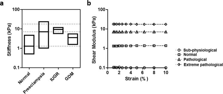
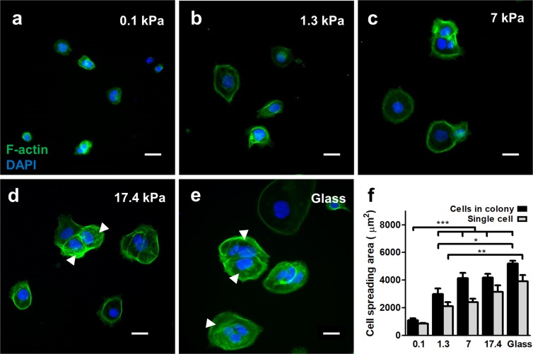
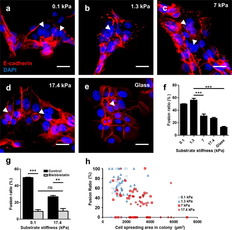
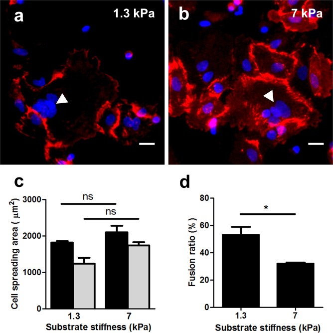
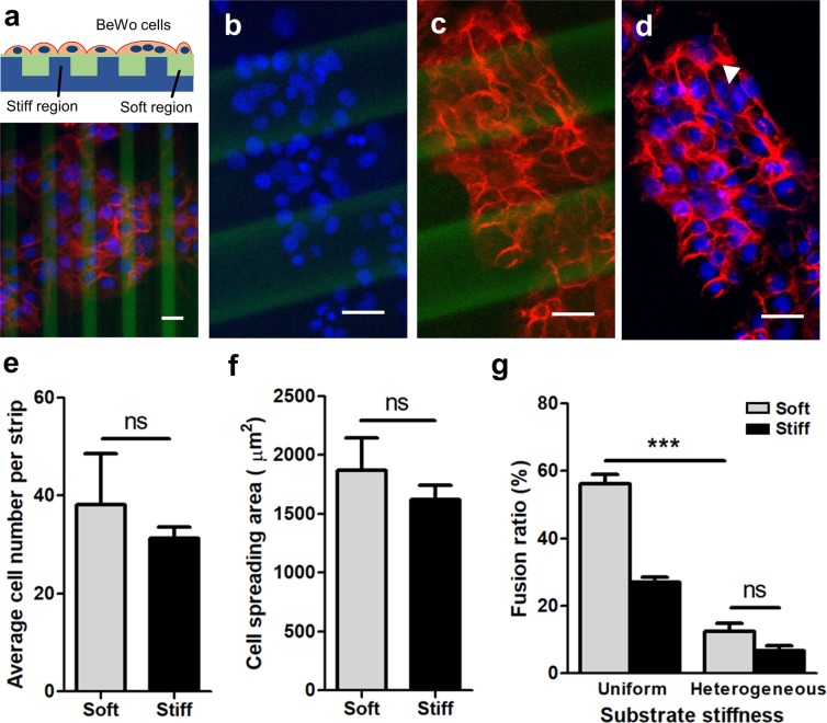
The syncytiotrophoblast is a multinucleated layer that plays a critical role in regulating functions of the human placenta during pregnancy. Maintaining the syncytiotrophoblast layer relies on ongoing fusion of mononuclear cytotrophoblasts throughout pregnancy, and errors in this fusion process are associated with complications such as preeclampsia. While biochemical factors are known to drive fusion, the role of disease-specific extracellular biophysical cues remains undefined. Since substrate mechanics play a crucial role in several diseases, and preeclampsia is associated with placental stiffening, we hypothesize that trophoblast fusion is mechanically regulated by substrate stiffness. We developed stiffness-tunable polyacrylamide substrate formulations that match the linear elasticity of placental tissue in normal and disease conditions, and evaluated trophoblast morphology, fusion, and function on these surfaces. Our results demonstrate that morphology, fusion, and hormone release is mechanically-regulated via myosin-II; optimal on substrates that match healthy placental tissue stiffness; and dysregulated on disease-like and supraphysiologically-stiff substrates. We further demonstrate that stiff regions in heterogeneous substrates provide dominant physical cues that inhibit fusion, suggesting that even focal tissue stiffening limits widespread trophoblast fusion and tissue function. These results confirm that mechanical microenvironmental cues influence fusion in the placenta, provide critical information needed to engineer better in vitro models for placental disease, and may ultimately be used to develop novel mechanically-mediated therapeutic strategies to resolve fusion-related disorders during pregnancy.


Conflict of interest statement

The authors declare no competing interests.

Figures

Figure 1
Figure 1
Design criteria and mechanical properties for stiffness-tunable substrates. (a) Tissue stiffness of normal and diseased placenta (log scale, shear modulus values), using data obtained from literature. (b) Measured shear modulus of synthesized polyacrylamide hydrogels measured over an applied strain of 10%. Loss modulus measurements were negligible.
Figure 2
Figure 2
Effects of substrate stiffness on BeWo cell morphology. (ae) Representative figures of BeWo cells cultured on substrates with (a) 0.1 kPa, (b) 1.3 kPa, (c) 7 kPa, (d) 17.4 kPa, and (e) glass substrates. Green: f-actin; blue: nuclear DAPI. Arrows indicate stress fibers. Scale bar is 50 μm. (f) Measured average cell spread area of BeWo cells cultured on various substrates. (data reported as mean ± standard deviation for n = 3 independent experiments; *p < 0.05; **p < 0.01; ***p < 0.001, by two-tailed, one-way ANOVA with Holm-Sidak Post-hoc comparison).
Figure 3
Figure 3
Effects of substrate stiffness on BeWo cell fusion. (ae) Representative figures of syncytium formed on substrates with (a) 0.1 kPa, (b) 1.3 kPa, (c) 7 kPa, (d) 17.4 kPa, and (e) glass substrates. Red: E-cadherin; blue: DAPI. Arrowheads indicate syncytial regions. Scale bar is 50 μm. (f) BeWo cell fusion ratio was greatly enhanced on substrate matching normal placental tissue stiffness. (g) Fusion was greatly suppressed after blebbistatin inhibition for cells cultured on ultrasoft and ultrastiff substrates. Data reported as mean ± standard deviation for n = 3 independent experiments; **p < 0.01, ***p < 0.001, by Student’s t-test. (h) No correlation was found between cell spreading area and fusion ratio for cells cultured on all the substrate stiffness tested.
Figure 4
Figure 4
Effects of substrate stiffness on BeWo cell β-hCG expression. (a,b) BeWo cells only express β-hCG with forskolin treatment. (c,d) Representative figures of cells expressing β-hCG on soft (c) and stiff (d) substrates. Red: β-hCG; blue: DAPI. Scale bar is 50 μm. (e) Ratio of BeWo cells expressing β-hCG and (f) normalized media β-hCG level when cultured on substrates with various stiffness. Data reported as mean ± standard deviation for n = 3 independent experiments; *p < 0.05, **p < 0.01, ***p < 0.001, by two-tailed, one-way ANOVA with Holm-Sidak Post-hoc comparison.
Figure 5
Figure 5
Effects of substrate stiffness on vCTBs morphology and fusion. (a,b) Representative figures of vCTBs cultured on substrates with normal (a) and preeclamptic (b) tissue stiffness after 72-hour primary culture. Red: E-cadherin; blue: DAPI. Scale bar is 50 μm. (c) No significant difference was observed for vCTBs cultured on different substrates stiffness tested. (d) Fusion ratio was greatly enhanced for vCTBs cultured on substrate matching normal placental tissue stiffness. Data reported as mean ± standard deviation for n = 3 independent experiments; *p < 0.05, by Student’s t-test.
Figure 6
Figure 6
Effects of heterogeneous substrate stiffness on BeWo cell fusion. (a) Schematic of the mechanically patterned composite hydrogel for cell culture (top) and the representative figures of BeWo cells cultured after 48-hour forskolin induction. (b–d) Representative images of (b) nuclei, (c) E-cadherin, and (d) merged on heterogeneously stiff substrates. Red: E-cadherin; blue: DAPI; green: fluorescein. Scale bar is 50 μm. (e,f) No significant difference was observed regarding (e) average cell number per strip and (f) cell spreading area. (g) BeWo cell fusion ratio was greatly suppressed when cultured on heterogeneous substrate with stiff “hot spots”. Data reported as mean ± standard deviation for n = 3 independent experiments; ***p < 0.001, by Student’s t-test.

Similar articles

Cited by

References

    1. Gude NM, Roberts CT, Kalionis B, King RG. Growth and function of the normal human placenta. Thrombosis Res. 2004;114:397–407. doi: 10.1016/j.thromres.2004.06.038. - DOI - PubMed
    1. Fuchs R, Ellinger I. Endocytic and Transcytotic Processes in Villous Syncytiotrophoblast: Role in Nutrient Transport to the Human Fetus. Traffic. 2004;5:725–738. doi: 10.1111/j.1600-0854.2004.00221.x. - DOI - PubMed
    1. Thellin O. Pregnancy and the immune system: between tolerance and rejection. Toxicology. 2003;185:179–184. doi: 10.1016/S0300-483X(02)00607-8. - DOI - PubMed
    1. Morgan TK. Role of the Placenta in Preterm Birth: A Review. Am. J. Perinatol. 2016;33:258–266. doi: 10.1055/s-0035-1570379. - DOI - PubMed
    1. Law A, et al. The prevalence of complications and healthcare costs during pregnancy. J. Med. Econ. 2015;18:533–541. doi: 10.3111/13696998.2015.1016229. - DOI - PubMed

Publication types