

Transcriptomic features of tumour-infiltrating CD4lowCD8high double positive αβ T cells in melanoma
- PMID: 32246006
- PMCID: PMC7125144
- DOI: 10.1038/s41598-020-62664-x
Transcriptomic features of tumour-infiltrating CD4lowCD8high double positive αβ T cells in melanoma
Abstract
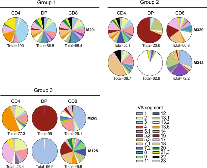
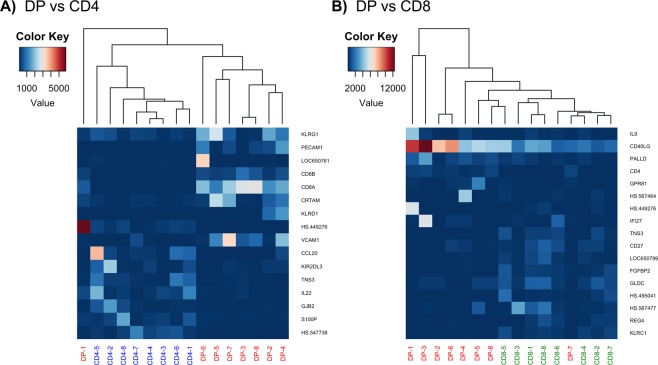
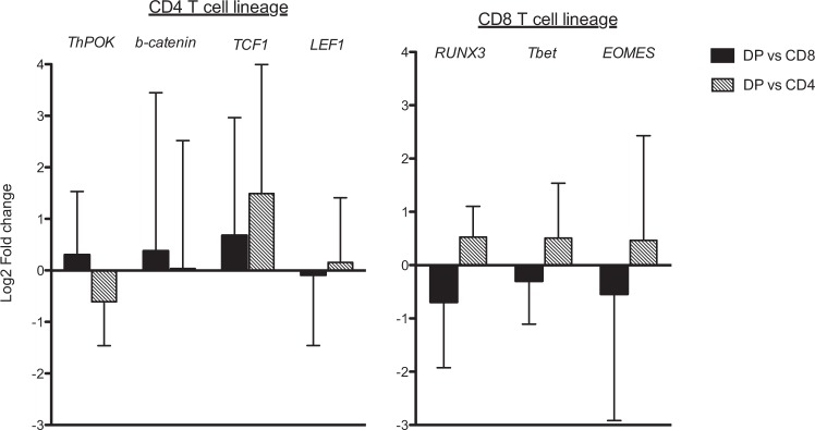
Peripheral CD4+CD8+ double positive (DP) T cells are a phenotypically and functionally heterogeneous population depending on their origin and pathologic context. We previously identified among tumour infiltrating lymphocytes in melanoma, a tumour-reactive MHC class-I restricted CD4lowCD8high DP αβ T-cell subpopulation with CD4-like function. In this study, we used an in-depth comparative transriptomic analysis of intra-melanoma DP T cells and CD4 and CD8 single positive (SP) T cells, to better comprehend the origin of this DP phenotype, and define the transcriptomic signature of activated DP T cells. We observed that intra-melanoma DP T cells were transcriptome-wise closer to their CD8 SP T-cell counterparts in terms of number of genes differentially expressed (97 in common with CD8 SP T cells and 15 with CD4 SP T cells) but presented hallmarks of a transition to a CD4-like functional profile (CD40LG) with a decreased cytotoxic signature (KLRC1) in favour of an increased cytokine-receptor interaction signature (IL4, IL24, IL17A…). This unleashed CD4-like program could be the results of the observed unbalanced expression of the THPOK/Runx3 transcription factors in DP T cells. Overall, this study allow us to speculate that intra-melanoma DP T cells arise from CD8 SP T cells being reprogrammed to a helper function.
Conflict of interest statement
The authors declare no competing interests.
Figures
References
-
- Blue ML, Daley JF, Levine H, Schlossman SF. Coexpression of T4 and T8 on peripheral blood T cells demonstrated by two-color fluorescence flow cytometry. The Journal of Immunology. 1985;134:2281–2286. - PubMed
-
- Cortés KC, et al. Expression of programmed cell death protein 1 and T-cell immunoglobulin- and mucin-domain-containing molecule-3 on peripheral blood CD4+CD8+ double positive T cells in patients with chronic hepatitis C virus infection and in subjects who spontaneously cleared the virus. Journal of Viral Hepatitis. 2019;26:942–950. - PMC - PubMed
Publication types
MeSH terms
Substances
LinkOut - more resources
Full Text Sources
Medical
Molecular Biology Databases
Research Materials
