

Renoprotective and neuroprotective effects of enteric hydrogen generation from Si-based agent
- PMID: 32246095
- PMCID: PMC7125117
- DOI: 10.1038/s41598-020-62755-9
Renoprotective and neuroprotective effects of enteric hydrogen generation from Si-based agent
Abstract
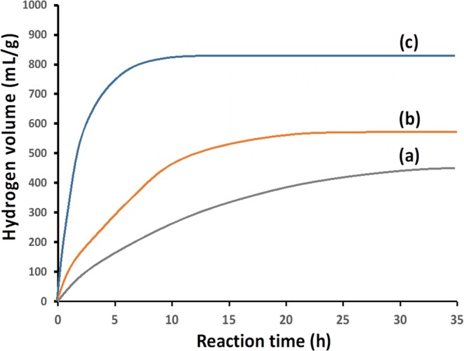
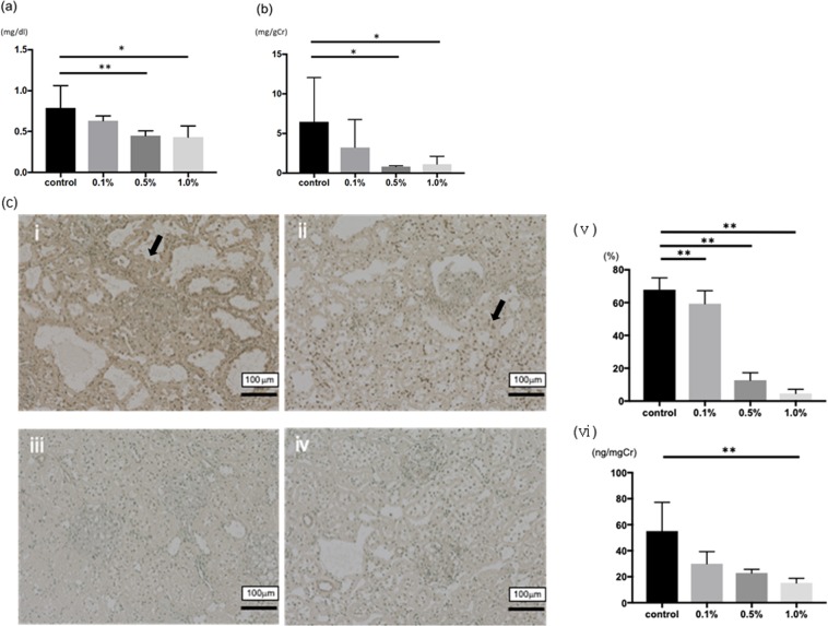
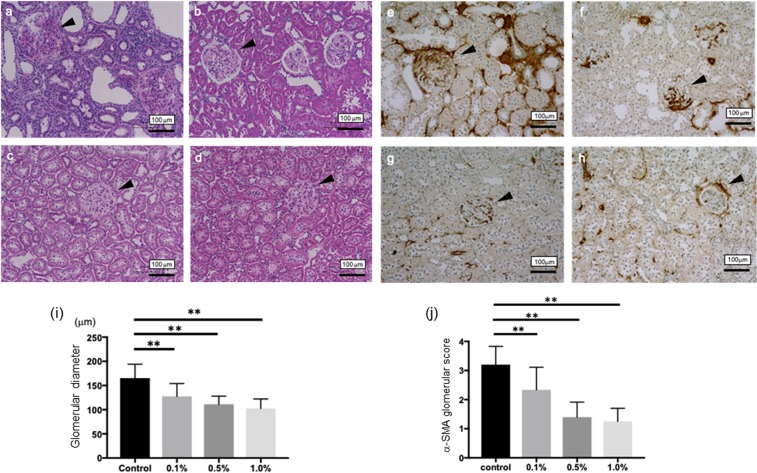
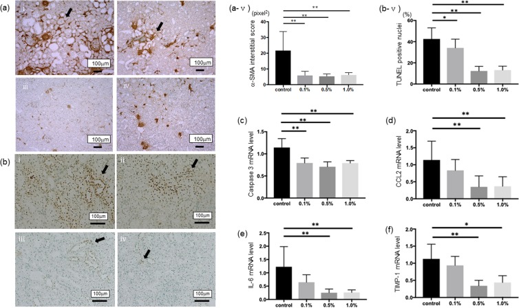
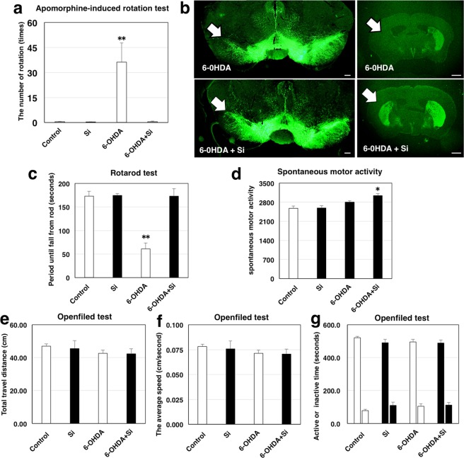
We have developed Si-based agent which can generate a large amount of hydrogen. Si-based agent continues generating hydrogen for more than 24 h by the reaction with water under conditions similar to those in bowels, i.e., pH8.3 and 36 °C, and generates ~400 mL hydrogen. To investigate beneficial effects for diseases associated with oxidative stress, Si-based agent is administered to remnant kidney rats and Parkinson's disease mice. Rats are fed with control or Si-based agent-containing diet for 8 weeks. Si-based agent is found to greatly suppress the development of renal failure and the parameters of oxidative stress. Treatment with Si-based agent in a mouse model of hemi-Parkinson's disease induced by 6-hydroxydopamine attenuated degeneration of dopaminergic neurons and prevented impairment of motor balance and coordination. These findings indicate that the Si-based agent shows renoprotective and neuroprotective effects presumably via suppression of oxidative stress by generation of hydrogen.
Conflict of interest statement
The authors declare no competing interests.
Figures
Similar articles
-
Diverse Possibilities of Si-Based Agent, a Unique New Antioxidant.Antioxidants (Basel). 2023 May 8;12(5):1061. doi: 10.3390/antiox12051061. Antioxidants (Basel). 2023. PMID: 37237927 Free PMC article. Review.
-
Efficacy of a Si-based agent against developing renal failure in a rat remnant kidney model.Biochem Biophys Res Commun. 2020 Dec 17;533(4):698-703. doi: 10.1016/j.bbrc.2020.10.067. Epub 2020 Oct 31. Biochem Biophys Res Commun. 2020. PMID: 33131768
-
Neuroprotective effects of Si-based hydrogen-producing agent on 6-hydroxydopamine-induced neurotoxicity in juvenile mouse model.Behav Brain Res. 2024 Jun 25;468:115040. doi: 10.1016/j.bbr.2024.115040. Epub 2024 May 7. Behav Brain Res. 2024. PMID: 38723675
-
Evaluation of the antiparkinsonism and neuroprotective effects of hydrogen sulfide in acute 6-hydroxydopamine-induced animal model of Parkinson's disease: behavioral, histological and biochemical studies.Neurol Res. 2018 Jul;40(7):523-531. doi: 10.1080/01616412.2017.1390903. Epub 2018 May 4. Neurol Res. 2018. PMID: 29726751
-
Leads for the development of neuroprotective treatment in Parkinson's disease and brain imaging methods for estimating treatment efficacy.Eur J Pharmacol. 1999 Jun 30;375(1-3):75-86. doi: 10.1016/s0014-2999(99)00260-5. Eur J Pharmacol. 1999. PMID: 10443566 Review.
Cited by
-
Small Intestinal Bacterial Overgrowth as Potential Therapeutic Target in Parkinson's Disease.Int J Mol Sci. 2021 Oct 28;22(21):11663. doi: 10.3390/ijms222111663. Int J Mol Sci. 2021. PMID: 34769091 Free PMC article. Review.
-
Molecular Hydrogen Therapy-A Review on Clinical Studies and Outcomes.Molecules. 2023 Nov 26;28(23):7785. doi: 10.3390/molecules28237785. Molecules. 2023. PMID: 38067515 Free PMC article. Review.
-
Nutrition and Cancer Risk from the Viewpoint of the Intestinal Microbiome.Nutrients. 2021 Sep 23;13(10):3326. doi: 10.3390/nu13103326. Nutrients. 2021. PMID: 34684330 Free PMC article. Review.
-
Diverse Possibilities of Si-Based Agent, a Unique New Antioxidant.Antioxidants (Basel). 2023 May 8;12(5):1061. doi: 10.3390/antiox12051061. Antioxidants (Basel). 2023. PMID: 37237927 Free PMC article. Review.
-
Efficacy of the silicon based agent for age related decline in vestibular function.Sci Rep. 2025 Aug 14;15(1):29790. doi: 10.1038/s41598-025-14302-7. Sci Rep. 2025. PMID: 40813876 Free PMC article.
References
Publication types
MeSH terms
Substances
LinkOut - more resources
Full Text Sources
Other Literature Sources
Medical
