. 2020 Jul;38(7):890-903.
doi: 10.1002/stem.3181. Epub 2020 Apr 30.

Lin28 reprograms inner ear glia to a neuronal fate

Affiliations

Lin28 reprograms inner ear glia to a neuronal fate

Judith S Kempfle et al. Stem Cells. 2020 Jul.

Abstract

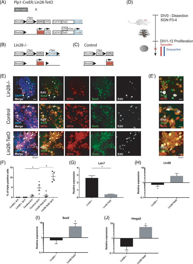
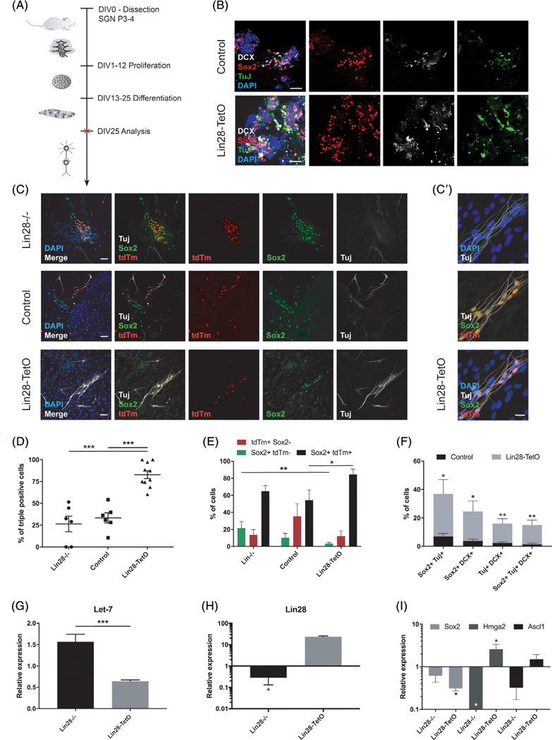
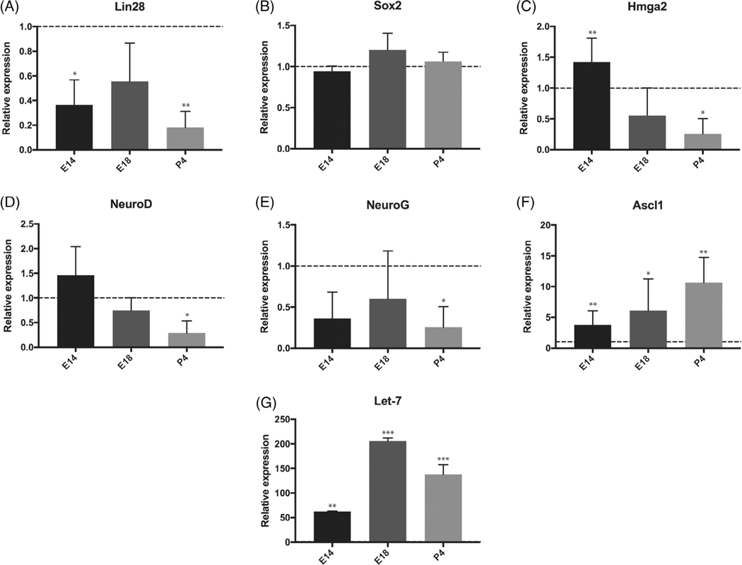
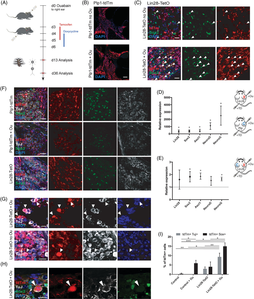
Sensorineural hearing loss is irreversible and can be caused by loss of auditory neurons. Regeneration of neural cells from endogenous cells may offer a future tool to restore the auditory circuit and to enhance the performance of implantable hearing devices. Neurons and glial cells in the peripheral nervous system are closely related and originate from a common progenitor. Prior work in our lab indicated that in the early postnatal mouse inner ear, proteolipid protein 1 (Plp1) expressing glial cells could act as progenitor cells for neurons in vitro. Here, we used a transgenic mouse model to transiently overexpress Lin28, a neural stem cell regulator, in Plp1-positive glial cells. Lin28 promoted proliferation and conversion of auditory glial cells into neurons in vitro. To study the effects of Lin28 on endogenous glial cells after loss of auditory neurons in vivo, we produced a model of auditory neuropathy by selectively damaging auditory neurons with ouabain. After neural damage was confirmed by the auditory brainstem response, we briefly upregulated the Lin28 in Plp1-expressing inner ear glial cells. One month later, we analyzed the cochlea for neural marker expression by quantitative RT-PCR and immunohistochemistry. We found that transient Lin28 overexpression in Plp1-expressing glial cells induced expression of neural stem cell markers and subsequent conversion into neurons. This suggests the potential for inner ear glia to be converted into neurons as a regeneration therapy for neural replacement in auditory neuropathy.

Keywords: glial conversion; inner ear; mouse models; neural stem cells; regeneration.


Conflict of interest statement

CONFLICT OF INTEREST

The authors declared no potential conflicts of interest.

Figures

FIGURE 1
FIGURE 1
Proliferation of Plp1-expressing glial cells after knockout or overexpression of Lin28 in vitro. A, For Lin28 overexpression in Plp1-positive cells, Plp1-CreER;STOP fl; rtTA mice and Lin28a/b-TetO, Lafayette, Colorado; STOP fl; tdTm (tdTm) mice were crossed to obtain Plp1-CreER;rtTA;Lin28a/b-TetO;tdTm (Lin28-TetO) transgenic mice. Tamoxifen application initiated Cre recombinase dependent tdTm and tetracycline-controlled transactivator (rtTA) expression. In the presence of doxycycline, the tetracycline operator (TetO) was transiently activated and led to overexpression of Lin28. B, For the Lin28 knockout experiments, Plp1-CreER mice were crossed with Lin28a-fl;tdTm mice to obtain Plp1-CreER;Lin28a-fl;tdTm (Lin28−/−) transgenic mice. Application of tamoxifen led to conditional knockout of Lin28 in tdTm expressing Plp1 positive cells. C, Plp1-CreER;tdTm mice were used as controls for Lin28 overexpression and knockout experiments (control). D, Timeline for neurosphere assay from early postnatal spiral ganglion cells. Pups were dissected at P3-P4 and kept under proliferating conditions for 12 days, undergoing two passages; DIV, days in vitro. Spheres were treated after the second passage and analyzed by immunohistochemistry (red X). E, Second-generation neurospheres were treated with ethynyl-desoxyuridine (EdU) for either 6 or 24 hours and harvested for immunohistochemistry. Neurospheres of Lin28−/− (first row), control (second row), and Lin28-TetO (third row) mice were stained for EdU (white) and Sox2 (green), nuclei were labeled with DAPI (blue). Plp1-positive cells expressed tdTm (red). Merged images in the first two columns show co-localization. Scale bar = 20 μm. E′, High-magnification image of EdU-Sox2-tdTm colocalization. Scale bar = 10 μm. F, Quantification of tdTm, Sox2, and EdU-triple-positive (+) cells as percentage of the total number of tdTm+ cells after 6 or 24 hours of EdU treatment (*P < .05, mean ± SEM). G, Relative expression of Let-7 in proliferating Lin28−/− and Lin28-TetO neurospheres (mean ± SEM, *P < .05, n = 3). H-J, Quantitative RT-PCR of Lin28 (n = 7), Sox2 (n = 5), and Hmga2 (n = 6) in proliferating Lin28−/− and Lin28-TetO neurospheres compared to control (mean ± SEM, *P < .05, control set as 1)
FIGURE 2
FIGURE 2
Differentiation of Plp1-expressing glial cells after knockout or overexpression of Lin28 in vitro. A, Timeline for in vitro neurosphere assay from early postnatal spiral ganglion cells. Pups were dissected at P3-P4 and kept under proliferating conditions for 12 days, undergoing two passages; DIV, days in vitro. Spheres were treated after the second passage and analyzed by immunohistochemistry (red cross). B, Immunohistochemical analysis of third-generation spiral ganglion neurospheres after 12 days of differentiation. Differentiating neurospheres of Lin28-TetO and control mice were stained for DCX (white), Tuj (green), and Sox2 (red). Nuclei were labeled with DAPI (blue). Scale bar = 50 μm. C, Immunohistochemical analysis of third-generation spiral ganglion neurospheres after 12 days of differentiation. Differentiating neurospheres of Lin28−/− (first row), control (second row), and Lin28-TetO mice (third row) were stained for TuJ (white) and Sox2 (green), and colocalized with tdTm (red). Nuclei were labeled with DAPI (blue). Scale bar = 50 μm. C′, High-magnification of Lin28-TetO, Plp1-positive glial cells (tdTm) coexpressing Sox2 (green) and TuJ (white). Nuclei were labeled with DAPI (blue). Scale bar = 20 μm. D, Quantification of tdTm, Sox2, and TuJ positive (+) cells in differentiating neurospheres of Lin28−/−, control, and Lin28-TetO mice as percentage of the total number of tdTm+ Plp1 progenitors after 12 days of differentiation (***P < .001, mean ± SEM). E, Quantification of tdTm, Sox2 double positive, tdTm single positive, and Sox2 single positive cells in spheres on day 1 of differentiation (**P < .01, *P < .05, mean ± SEM, n = 3). F, Number of cells coexpressing Sox2, TuJ, and/or DCX per total cell number in differentiating neurospheres from control or Lin28-TetO mice at 12 days of differentiation (**P < .01, *P < .05, mean ± SEM, n = 3). G, Quantitative expression of Let-7 in differentiating spiral ganglion neurospheres after Lin28 deletion (Lin28−/−) or Lin28 overexpression (Lin28-TetO) (***P < .001, mean ± SEM, n = 3). H-I, Quantitative RT-PCR of Lin28, Sox2, Hmga2, and Ascl1 in differentiating Lin28−/− and Lin28-TetO neurospheres compared to control (*P < .05, mean ± SEM, n = 3, control set as 1)
FIGURE 3
FIGURE 3
Expression of Lin28, Let-7, and neural stem cell markers in the developing ear. A-F, RT-PCR in wild-type embryos from embryonic day E9 to P4. Expression level at E9 is set as 1 and serves as a control. Experimental values at E14, E18, and P4 are compared with E9 for each gene. Neural stem cell genes Lin28 and Hmga2 were downregulated between E9 and P4. The expression of early neural progenitor genes, Neurog1 (NeuroG) and Neurod1 (NeuroD), was decreased during maturation of SGNs, while expression of more mature neural precursor marker, Ascl1, was increased between E9 and P4. Sox2 expression was unchanged (*P < .05; **P < .01, mean ± SEM, n = 3). G, Let-7 expression was increased from E9 to P4 (**P < .01; ***P < .001, mean ± SEM, n = 3)
FIGURE 4
FIGURE 4
Neural conversion of adult spiral ganglion glia after damage and transient Lin28 overexpression. A, Timeline for in vivo experiment. Ouabain surgery was performed on 6-week-old Lin28-TetO mice or Plp1-tdTm mice at experimental time point day 0 (d0), followed by tamoxifen injections from day 3 to day 5 and doxycycline injections from day 4 to day 6 after surgery. Animals were analyzed 7 days after injection at experimental day 13 or 30 days after injections at experimental day 36. B, Low-magnification view of proliferating Plp1-positive glia in the modiolus of the cochlea without (no Ou) or with (+Ou) ouabain treatment. Plp1-positive glia are shown in red (tdTm reporter). Nuclei were labeled with DAPI (blue). Scale bar = 100 μm. C Coexpression (arrowheads) of tdTm reporter (red) and Sox2 (green) was increased after ouabain treatment and Lin28 overexpression (Lin28-TetO + Ou) compared to Lin28 overexpression alone (Lin28-TetO no Ou). Scale bar = 20 μm. D, Seven days post Lin28 upregulation after tamoxifen/doxycycline injection (+TD) and ouabain treatment (+Ou). RT-PCR showed increased expression of Lin28, Sox2, Ascl1, Neurod1 (NeuroD), and Neurog1 (NeuroG) in Lin28-TetO (Lin28, X) mice compared to tamoxifen/doxycycline injected Plp1-tdTm control (Plp, C) mice after ouabain treatment (*P < .05, mean ± SEM, n = 5). E, Seven days post Lin28 upregulation after tamoxifen/doxycycline injection (+TD) and ouabain treatment (+Ou), RT-PCR showed higher expression of Sox2, Ascl1, Neurod1 (NeuroD), and Neurog1 (NeuroG) in ouabain damaged right ears (Lin28, X) of Lin28-TetO mice compared to undamaged left ears (Lin28, C, set at 1) (*P < .05, mean ± SEM, n = 4). F, Low-magnification of Plp1-tdTm mice after tamoxifen injections with or without ouabain, showing no coexpression of Plp1-expressing glial cells (tdTm in red) and Tuj (white). Ouabain-treated and tamoxifen injected Plp1-tdTm mice (+ Ou) showed diminished Tuj expression. Plp1 glia were found with Sox2 coexpression (green), but not with Tuj (white). Uninjected Lin28-TetO mice without ouabain never demonstrated Tuj-expressing Plp1-glia. Nuclei were labeled with DAPI (blue). Scale bar = 25 μm. G, Multiple lineage-traced (white arrowheads, upper row) Plp1-positive glial cells (tdTm, red) 4 weeks after ouabain-induced neural damage (+Ou) and Lin28 upregulation via tamoxifen/doxycycline injections expressed TuJ (white). Lin28 upregulation without previous ouabain treatment (no Ou) resulted in less Plp1-positive cells that expressed TuJ (single white arrowhead, bottom row). Scale bar = 10 μm. H, Plp1-positive glial cell (tdTm, in red) 4 weeks after ouabain-induced neural damage and Lin28 upregulation via tamoxifen/doxycycline injections (white arrowhead) co-expressed TuJ (white) but had lost Sox2 (green) expression. Nuclei were labeled with DAPI (blue). Scale bar = 10 μm. I, Quantification of surviving lineage-traced Plp1-positive glial cells (tdTm) co-expressing TuJ after ouabain and Lin28 upregulation via tamoxifen/doxycycline injections, compared to Lin28 upregulation only without ouabain. Lin28 overexpression after ouabain led to higher numbers of converted glial cells than Lin28 upregulation without previous ouabain treatment (**P < .01, mean ± SEM, n = 7)

Similar articles

Cited by

References

    1. Nadol JB Jr, Young YS, Glynn RJ. Survival of spiral ganglion cells in profound sensorineural hearing loss: implications for cochlear implantation. Ann Otol Rhinol Laryngol. 1989;98(6):411–416. - PubMed
    1. Nadol JB Jr. Patterns of neural degeneration in the human cochlea and auditory nerve: implications for cochlear implantation. Otolaryngol Head Neck Surg. 1997;117(3 Pt 1):220–228. - PubMed
    1. Seyyedi M, Viana LM, Nadol JB Jr. Within-subject comparison of word recognition and spiral ganglion cell count in bilateral cochlear implant recipients. Otol Neurotol. 2014;35(8):1446–1450. - PMC - PubMed
    1. Moller AR. History of cochlear implants and auditory brainstem implants. Adv Otorhinolaryngol. 2006;64:1–10. - PubMed
    1. Lustig LR, Yeagle J, Driscoll CL, Blevins N, Francis H, Niparko JK. Cochlear implantation in patients with neurofibromatosis type 2 and bilateral vestibular schwannoma. Otol Neurotol. 2006;27(4):512–518. - PubMed

Publication types