Review
. 2020 Oct;15(10):1821-1830.
doi: 10.4103/1673-5374.280306.

Spinal genesis of Mayer waves

Affiliations
Review

Spinal genesis of Mayer waves

George Zaki Ghali et al. Neural Regen Res. 2020 Oct.

Retraction in

Abstract

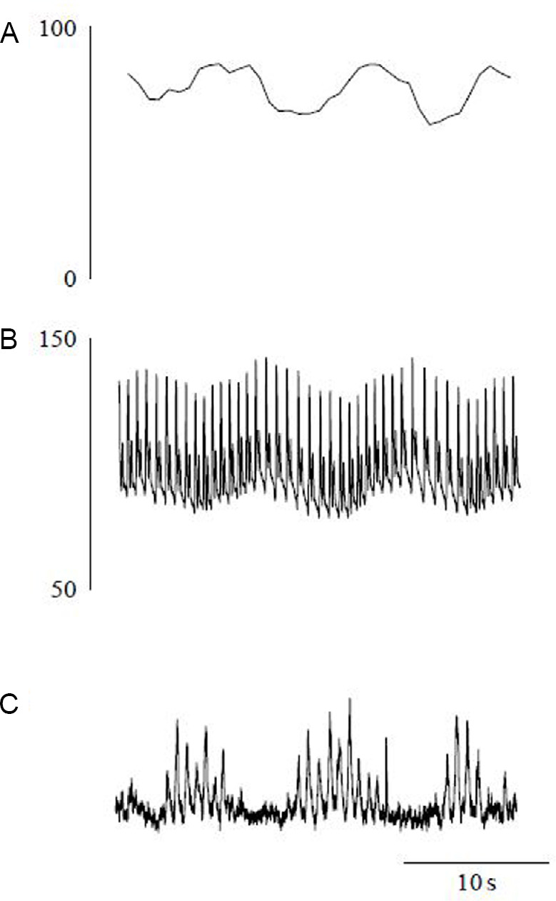
Variability in cardiovascular spectra was first described by Stephan Hales in 1733. Traube and Hering initially noted respirophasic variation of the arterial pressure waveform in 1865 and Sigmund Mayer noted a lower frequency oscillation of the same in anesthetized rabbits in 1876. Very low frequency oscillations were noted by Barcroft and Nisimaru in 1932, likely representing vasogenic autorhythmicity. While the origins of Traube Hering and very low frequency oscillatory variability in cardiovascular spectra are well described, genesis mechanisms and functional significance of Mayer waves remain in controversy. Various theories have posited baroreflex and central supraspinal mechanisms for genesis of Mayer waves. Several studies have demonstrated the persistence of Mayer waves following high cervical transection, indicating a spinal capacity for genesis of these oscillations. We suggest a general tendency for central sympathetic neurons to oscillate at the Mayer wave frequency, the presence of multiple Mayer wave oscillators throughout the brainstem and spinal cord, and possible contemporaneous genesis by baroreflex and vasomotor mechanisms.

Keywords: Mayer waves; central; cervical; genesis; origins; spinal cord; sympathogenesis; transection.


Conflict of interest statement

None

Figures

Figure 1
Figure 1
Effects of gravity upon cardiovascular variabilities in a healthy subject. (A) Heart rate. (B) Invasive arterial pressure. (C) Mean sympathetic nerve activity. The traces represent 30 second recordings taken starting during the 5th minute of a tilt maneuver gravitational stimulus. Modified with permission from of Montano et al. (2009a).
Figure 2
Figure 2
MAP and RSNA power spectra and coherence in conscious rats. MAP and RSNA recordings were performed contemporaneously in sham-operated (n = 10; heavy lines) and sinoaortic barodenervated (n = 10; thin lines) conscious rats. Fast Fourier transformation (34 periods of 204.8-second duration, representing 2048 data points, overlapping by half the recorded epoch) was utilized to make determinations of power spectral densities and coherence. Respective mean prior to spectral analysis was used to normalize individual RSNA data sets. MAP: Mean arterial pressure; n.u.: normalized units; RSNA: renal sympathetic nerve activity. Modified with permission from Figure 1 of Julien (2006).
Figure 3
Figure 3
Recovery of arterial pressure recovery following high cervical (C1) transection. Femoral arterial pressure traces recovery of arterial pressure recovery following C1 transection Femoral arterial pressure traces (y axis; mmHg) in an unanesthetized decerebrate rat 10 minutes prior to spinalization (t = –10 minutes) in the absence of anesthesia, 5 minutes prior to spinalization (t = –5 minutes) in the presence of 2% isoflurane anesthesia (note depressor response), immediately following spinalization (t = 0 minute) (right upper subpanel), at which point isoflurane was turned off and at t = 20, 50, 80, 110, 140, and 170 minutes following spinalization. Timescale bar is 10 seconds. Modified with permission from Figure 2 of Ghali (2019).
Figure 4
Figure 4
Cardiovascular and respiratory variabilities in a vagotomized unanesthetized midcollicular decerebrate cat. Sympathetic nerve (SNA), electrocardiographic (ECG), arterial blood pressure (ABP), and ventilatory activity (VENTILATION) traces are demonstrated. Sympathetic activity and oscillatory rhythmicity of the cardiovascular variabilities persist following spinal transection and are augmented by aortic constriction mediated spinospinal sympathetic reflex. Modified with permission from Figure 1 of Montano et al. (2000).
Figure 5
Figure 5
Cervical transection and aortic constriction mediated changes in cardiovascular spectral variabilities in a vagotomized unanesthetized midcollicular decerebrate cat. Spectral analysis of the sympathetic nerve activity (SNA), cardiac interval (R-R interval), and systolic arterial pressure (SAP) evidences Mayer wave correspondent low frequency (LF) component and Traube Hering wave correspondent high frequency (HF) component. The ventilatory frequency evidences the Traube Hering correspondent high frequency component exclusively. Traube Hering and Mayer wave correspondent spectral peaks persist in all cardiovascular variabilities following cervical transection. Amplitude of sympathetic nerve activity and arterial pressure Traube Hering and Mayer waves is significantly increased in response to aortic constriction mediated spinospinal sympathetic reflex. The results collectively evidence the native capacity of the spinal cord to generate Mayer waves with oscillatory properties subject to peripheral modulation. Modified with permission from Figure 2 of Montano et al. (2000).

Similar articles

Cited by

References

    1. Accorsi-Mendonça D, da Silva MP, Souza GM, Lima-Silveira L, Karlen-Amarante M, Amorim MR, Almado CE, Moraes DJ, Machado BH. Pacemaking property of RVLM presympathetic neurons. Front Physiol. 2016;7:424. - PMC - PubMed
    1. Anderson B, Kenny BA, Neil E. Role of the chemoreceptors of the carotid and aortic regions in the production of the Mayer waves. Acta Physiol Scand. 1950;20:203. - PubMed
    1. Aoki M, Mori S, Kawahara K, Watanabe H, Ebata N. Generation of spontaneous respiratory rhythm in high spinal cats. Brain Res. 1980;202:51–63. - PubMed
    1. Baldridge BR, Burgess DE, Zimmerman EE, Carroll JJ, Sprinkle AG, Speakman RO, Li SG, Brown DR, Taylor RF, Dworkin S, Randall DC. Heart rate-arterial blood pressure relationship in conscious rat before vs. after spinal cord transection. Am J Physiol Regul Integr Comp Physiol. 2002;283:R748–756. - PubMed
    1. Barcroft J, Nisimaru Y. Rhythmical contraction of the spleen. J Physiol. 1932;74:294–298. - PMC - PubMed