

Pulmonary High-Resolution Computed Tomography (HRCT) Findings of Patients with Early-Stage Coronavirus Disease 2019 (COVID-19) in Hangzhou, China
- PMID: 32246819
- PMCID: PMC7156878
- DOI: 10.12659/MSM.923885
Pulmonary High-Resolution Computed Tomography (HRCT) Findings of Patients with Early-Stage Coronavirus Disease 2019 (COVID-19) in Hangzhou, China
Abstract
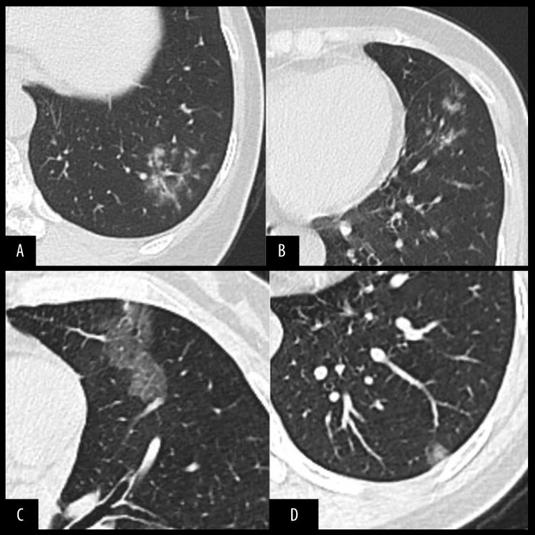
BACKGROUND The aim of this study was to investigate the imaging manifestations of early-stage coronavirus disease 2019 (COVID-19) and to provide imaging basis for early detection of suspected cases and stratified intervention. MATERIAL AND METHODS From 20 January 2020 to 2 February 2020, 6 patients diagnosed with COVID-19, including 1 male and 5 females, were retrospectively reviewed in Zhejiang Hospital. These cases were clinically assessed and classified as common COVID-19. All patients underwent thoracic high-resolution computed tomography (HRCT) within 2 days after the onset of symptoms, and their images were viewed by 2 radiologists who were blind to their clinical records. RESULTS CT images of 6 confirmed patients were collected. Two of the 6 patients (33.3%) had bilateral lung involvements and 4 (66.7%) had single-lung involvement. Two cases (33.3%) had a single lesion, 2 cases (33.3%) had 2 lesions, and 2 cases (33.3%) had multiple lesions. There were 2 cases (33.3%) with focal subpleural distribution and 1 case (16.7%) along the bronchial vascular bundle. Five cases (83.3%) had ground-glass opacities, 4 cases (66.7%) had ground-glass nodules, 1 case (16.7%) had thickened lobular septum, 2 cases (33.3%) had thickened bronchial wall, 2 cases (33.3%) had halo sign,1 case (16.7%) had crazy-paving sign, and 1 case (16.7%) had tree-in-bud sign. CONCLUSIONS The imaging manifestations of early-stage COVID-19 are relatively mild, and the imaging findings of some patients are not typical, which can easily lead to missed diagnoses. Thus, suspected cases need to be closely monitored, and epidemiological history and clinical laboratory examination should also be considered during diagnosis.
Figures
Similar articles
-
[Clinical features and high resolution CT imaging evolution of coronavirus disease 2019].Zhonghua Jie He He Hu Xi Za Zhi. 2020 Jun 12;43(6):509-515. doi: 10.3760/cma.j.cn112147-20200214-00094. Zhonghua Jie He He Hu Xi Za Zhi. 2020. PMID: 32486557 Chinese.
-
Interpretation of CT signs of 2019 novel coronavirus (COVID-19) pneumonia.Eur Radiol. 2020 Oct;30(10):5455-5462. doi: 10.1007/s00330-020-06915-5. Epub 2020 May 4. Eur Radiol. 2020. PMID: 32367422 Free PMC article.
-
CT imaging features of COVID-19 pneumonia: initial experience from Turkey.Diagn Interv Radiol. 2020 Jul;26(4):308-314. doi: 10.5152/dir.2020.20307. Diagn Interv Radiol. 2020. PMID: 32558645 Free PMC article.
-
[Diagnostic imaging findings in COVID-19].Ugeskr Laeger. 2020 Apr 6;182(15):V03200191. Ugeskr Laeger. 2020. PMID: 32286216 Review. Danish.
-
Thoracic imaging tests for the diagnosis of COVID-19.Cochrane Database Syst Rev. 2020 Sep 30;9:CD013639. doi: 10.1002/14651858.CD013639.pub2. Cochrane Database Syst Rev. 2020. Update in: Cochrane Database Syst Rev. 2020 Nov 26;11:CD013639. doi: 10.1002/14651858.CD013639.pub3. PMID: 32997361 Updated.
Cited by
-
High-Resolution CT Chest Findings in Suspected COVID-19 Pneumonia Patients With Negative Real-Time Polymerase Chain Reaction Assay.Cureus. 2021 Mar 21;13(3):e14023. doi: 10.7759/cureus.14023. Cureus. 2021. PMID: 33889463 Free PMC article.
-
Role of High Resolution Computed Tomography chest in the diagnosis and evaluation of COVID -19 patients -A systematic review and meta-analysis.Eur J Radiol Open. 2021;8:100350. doi: 10.1016/j.ejro.2021.100350. Epub 2021 May 13. Eur J Radiol Open. 2021. PMID: 34007865 Free PMC article.
-
Computed Tomography Severity Scoring on High-Resolution Computed Tomography Thorax and Inflammatory Markers With COVID-19 Related Mortality in a Designated COVID Hospital.Cureus. 2022 Apr 16;14(4):e24190. doi: 10.7759/cureus.24190. eCollection 2022 Apr. Cureus. 2022. PMID: 35592193 Free PMC article.
-
Computed Tomography (CT) Imaging Features of Patients with COVID-19: Systematic Review and Meta-Analysis.Radiol Res Pract. 2020 Jul 23;2020:1023506. doi: 10.1155/2020/1023506. eCollection 2020. Radiol Res Pract. 2020. PMID: 32733706 Free PMC article. Review.
-
Covid-19 imaging: A narrative review.Ann Med Surg (Lond). 2021 Sep;69:102489. doi: 10.1016/j.amsu.2021.102489. Epub 2021 Jun 18. Ann Med Surg (Lond). 2021. PMID: 34178312 Free PMC article. Review.
