Review
. 2020 Jul;190(7):1356-1369.
doi: 10.1016/j.ajpath.2020.03.007. Epub 2020 Apr 1.

The Pathobiology of Skin Aging: New Insights into an Old Dilemma

Affiliations
Review

The Pathobiology of Skin Aging: New Insights into an Old Dilemma

Eleanor Russell-Goldman et al. Am J Pathol. 2020 Jul.

Abstract

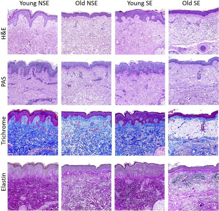
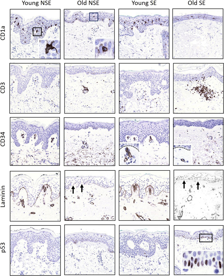
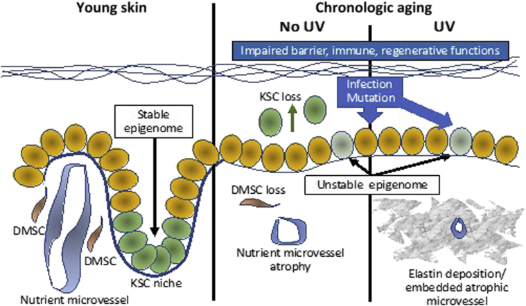
Long considered both physiologic and inevitable, skin aging is a degenerative phenomenon whereby both intrinsic and environmental factors conspire to produce an authentic disease. The consequences of this disorder are many and varied, ranging from atrophy and fragility to defective repair to deficient immunity and vulnerability to certain infections. The pathobiologic basis for skin aging remains poorly understood. At a cellular level, stem cell dysfunction and attrition appear to be key events, and both genetic and epigenetic factors are involved in a complex interplay that over time results in deterioration of our main protective interface with the external environment. Past and current understanding of the cellular and molecular intricacies of skin aging provide a foundation for future approaches designed to thwart the aging phenotype. Herein, the authors provide a review of current insights into skin aging, including the mechanisms of skin aging, the role of stem cells in skin aging and the implications of skin aging for the microbiome and for the development of cancer. Conquest of the oft overlooked disease of skin aging should have broad implications that transcend the integument and inform novel approaches to retarding aging and age-related dysfunction in those internal organs that youthful skin was designed to envelop and safeguard.


Figures

Figure 1
Figure 1
Histologic and histochemical survey of relatively young adult skin (29 years old) from face [chronically sun-exposed (SE)] and abdomen chronically non–sun-exposed (NSE), and relatively old skin (84 and 88 years old). By hematoxylin and eosin (H&E) and periodic acid-Schiff (PAS) staining, there is striking epidermal thinning with loss of the rete ridges in old skin independent of site/chronic UV exposure. In addition, note that old skin, again independent of site/chronic UV exposure, is characterized by less dense, less intensely stained reticular dermal collagen. The diminution of dermal collagen density in old skin is further confirmed by trichrome and elastin stains, which also disclose significant replacement of the uppermost dermis by markedly abnormal elastic fibers (so-called solar elastosis) only in the chronically sun-exposed facial skin, but not in the more photo-protected abdominal skin, of the older individual. Original magnification, ×10.
Figure 2
Figure 2
Immunohistochemical overview of sun-exposed (SE) and non–sun-exposed (NSE) skin from young and old patients characterized in Figure 1. Note depletion of CD1a-expressing epidermal Langerhans cells in relationship to age (independent of sun exposure) and altered cytology from more dendritic to rounded contours (boxed areas, shown in higher magnification in the insets). Sun-exposed old skin tends to harbor more CD3-expressing T cells, consistent with previously described heliodermatitis (Aging and Skin Immune Cells), and CD34 and laminin stains confirm loss of capillary loops (arrows) and superficial dermal microvascular density as a function of age. Dashed lines highlight the basement membrane with papillary dermal capillary loops below highlighted by CD34 immunostain (insets). Finally, discrete zones of p53 staining, consistent with mutant forms, are observed only in aged, sun-exposed epidermis, predominantly in the basal cell layer (boxed area). Original magnification, ×10 (main images); ×40 (insets).
Figure 3
Figure 3
Schematic summary of presumed alterations in skin stem cells with aging. Normal young skin with a stable epigenome maintains keratinocyte stem cells (KSCs) in niches at the tips of dermal papillae and in a microenvironment nourished by dermal papillary microvessels surrounded by dermal mesenchymal stem cells (DMSCs). With chronological aging, epigenomic instability, coupled with UV and environmentally-induced insults, induce mutations, compromise barrier function, and deplete the dermal microenvironment of nutrient vessels and DMSC, resulting in vulnerability to infection and neoplastic transformation. KSCs also become depleted, contributing to epidermal atrophy as a result of loss of the hemidesmosome component, collagen XVII (COL17A1), culminating in stem cell delamination and transepidermal elimination, rather than homeostatic maintenance.

References

    1. Scully J.L. What is a disease? EMBO Rep. 2004;5:650–653. - PMC - PubMed
    1. Makrantonaki E., Zouboulis C.C., German National Genome Research Network 2 The skin as a mirror of the aging process in the human organism--state of the art and results of the aging research in the German National Genome Research Network 2 (NGFN-2) Exp Gerontol. 2007;42:879–886. - PubMed
    1. Lener T., Moll P.R., Rinnerthaler M., Bauer J., Aberger F., Richter K. Expression profiling of aging in the human skin. Exp Gerontol. 2006;41:387–397. - PubMed
    1. Makrantonaki E., Brink T.C., Zampeli V., Elewa R.M., Mlody B., Hossini A.M., Hermes B., Krause U., Knolle J., Abdallah M., Adjaye J., Zouboulis C.C. Identification of biomarkers of human skin ageing in both genders. Wnt signalling - a label of skin ageing? PLoS One. 2012;7:1–10. - PMC - PubMed
    1. Kimball A.B., Alora-Palli M.B., Tamura M., Mullins L.A., Soh C., Binder R.L., Houston N.A., Conley E.D., Tung J.Y., Annunziata N.E., Bascom C.C., Isfort R.J., Jarrold B.B., Kainkaryam R., Rocchetta H.L., Swift D.D., Tiesman J.P., Toyama K., Xu J., Yan X., Osborne R. Age-induced and photoinduced changes in gene expression profiles in facial skin of Caucasian females across 6 decades of age. J Am Acad Dermatol. 2018;78:29–39.e7. - PubMed

Publication types