Review
. 2020:252:169-216.
doi: 10.1016/bs.pbr.2019.10.006. Epub 2019 Dec 5.

Innate and adaptive immune responses in Parkinson's disease

Affiliations
Review

Innate and adaptive immune responses in Parkinson's disease

Aubrey M Schonhoff et al. Prog Brain Res. 2020.

Abstract

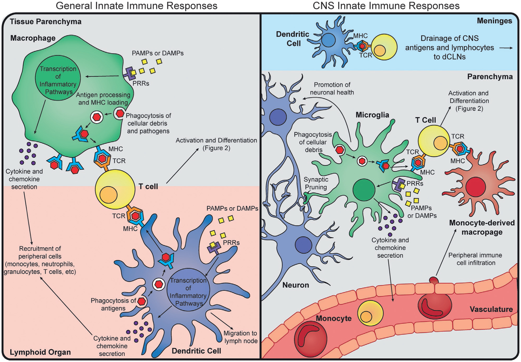
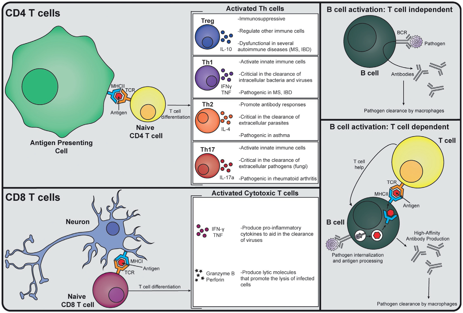
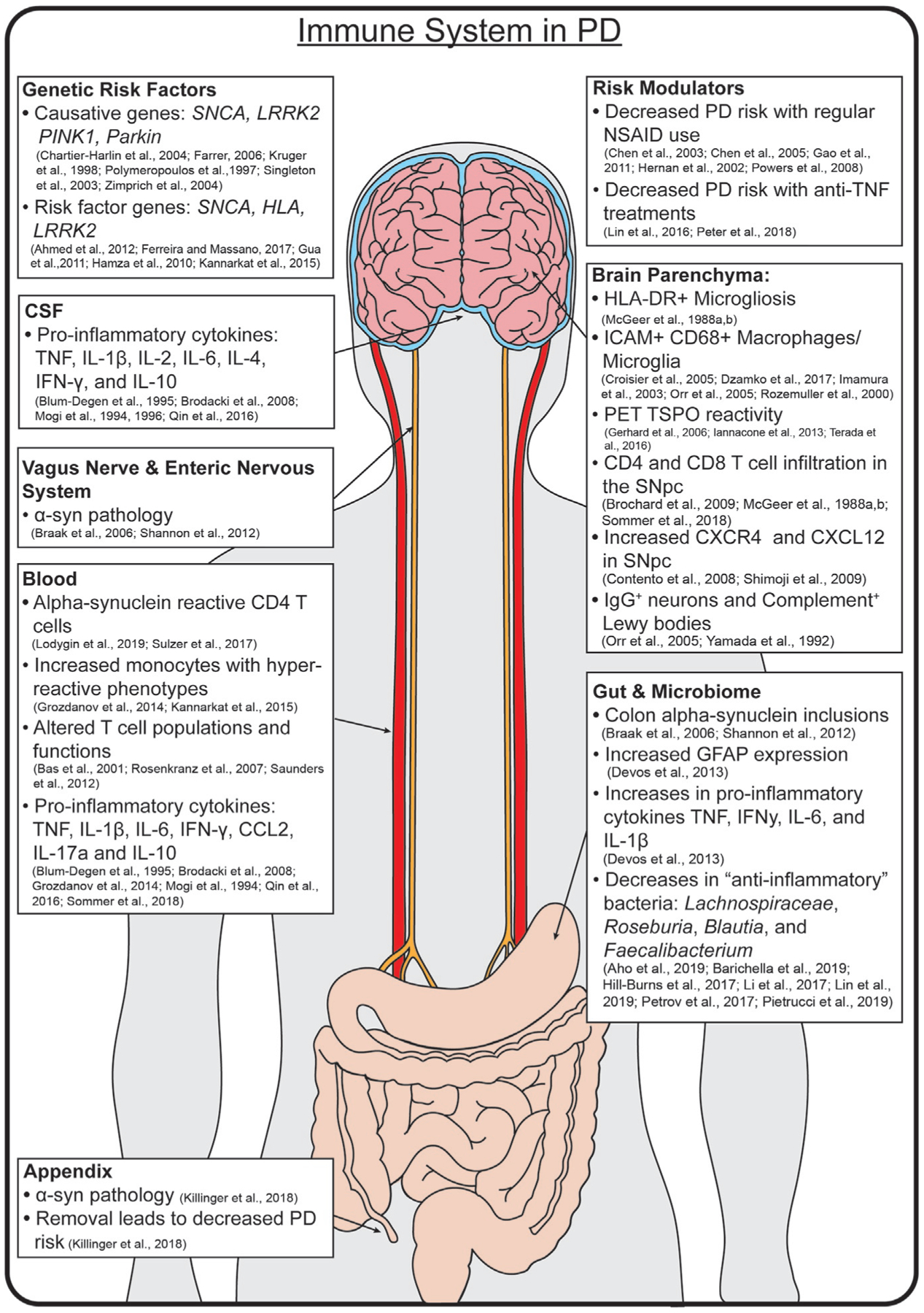
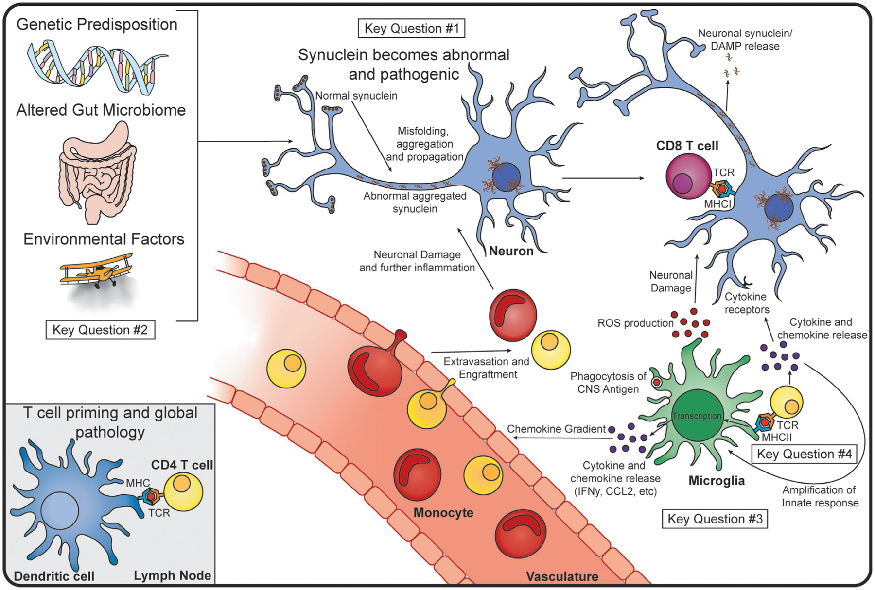
Parkinson's disease (PD) has classically been defined as a movement disorder, in which motor symptoms are explained by the aggregation of alpha-synuclein (α-syn) and subsequent death of dopaminergic neurons of the substantia nigra pars compacta (SNpc). More recently, the multisystem effects of the disease have been investigated, with the immune system being implicated in a number of these processes in the brain, the blood, and the gut. In this review, we highlight the dysfunctional immune system found in both human PD and animal models of the disease, and discuss how genetic risk factors and risk modifiers are associated with pro-inflammatory immune responses. Finally, we emphasize evidence that the immune response drives the pathogenesis and progression of PD, and discuss key questions that remain to be investigated in order to identify immunomodulatory therapies in PD.

Keywords: Alpha-synuclein; Immune system; Inflammation; Microbiota; Microglia; Parkinson's disease; T cells.


Figures

FIG. 1
FIG. 1
Overview of Innate Immune Responses. General innate immune responses typically involve antigen presenting cell (APC) recognition of pathogen associated molecular patterns (PAMPs, yellow squares) or danger associated molecular patterns (DAMPs) by a pattern recognition receptor (PRR, purple receptor). Upon recognition, the APC will undergo transcription for inflammatory pathways, which will lead to secretion of cytokines and chemokines (purple circles) to recruit more immune cells to the site, and upregulation of surface molecules involved in antigen presentation (MHC and costimulatory molecules, blue receptor). APCs can also phagocytose cellular debris and pathogens, process them, and load the antigenic peptides onto an MHC to be presented to T cells. Macrophages (green) typically carry out these functions within a tissue, whereas dendritic cells (blue) are usually found at tissue boundaries, and may migrate to a lymph node upon antigen uptake. In the CNS, the predominant APCs are microglia (green), although dendritic cells (blue) are found in the leptomeninges. Antigen uptake, pattern recognition, and cytokine secretion are thought to occur similar to general innate responses. Microglia also have tissue-specific homeostatic functions, such as synapse pruning and support of neuronal (blue) health. Infiltrating cells such as monocytes (red) and T cells (yellow), however, can be neuroprotective or neurotoxic, depending on the inflammatory stimulus. While there is some evidence that monocyte-derived macrophages (red) can play a role in antigen presentation during an inflammatory response, there is little known of their longevity in the parenchyma after resolution of the immune response.
FIG. 2
FIG. 2
Overview of Adaptive Immune Responses. CD4 T cells (yellow) interact with antigen presenting cells (APCs, green) via an antigen-loaded MHCII molecule, and costimulatory molecules (not shown). Upon antigen recognition by their T cell receptor (TCR), a CD4 T cell can differentiate into a Treg, Th1, Th2, or Th17 cell type, which have different roles in inflammation and produce signature cytokines. A CD8 T cell (maroon) interacts with antigen-loaded onto an MHCI molecule, which can be displayed on the surface of any cell, including neurons (blue). Upon antigen recognition by the TCR, a CD8 T cell will differentiate and produce inflammatory cytokines and lytic molecules. B cells (dark green) can act with or without the help of T cells. The B cell receptor (BCR) can recognize extracellular pathogens (purple), leading to the production and secretion of antibodies targeted toward that pathogen. A B cell can also recognize a pathogen via its BCR, process it, and load an antigenic peptide onto an MHCII molecule, which can then interact with the TCR of a CD4 T cell (yellow). This results in the production of high affinity antibodies targeted toward the pathogen that will promote that pathogen’s clearance.
FIG. 3
FIG. 3
The Immune System in Human PD. PD is a disease of global dysfunction, with inflammation found in multiple tissues. Risk is conferred by specific genes, and modulated by certain anti-inflammatory treatments. Notable inflammatory markers are found throughout the brain, CSF, blood, gut, enteric nervous system, and microbiome.
FIG. 4
FIG. 4
Proposed Mechanism of Immune Involvement in PD and Key Questions. We hypothesize that PD begins due to genetic predispositions, alterations in the gut microbiome, and the influence of external, environmental factors. It is possible that a CNS antigen drains to the lymph node (bottom left), where a dendritic cell (blue) can present it to a CD4 T cell (yellow), priming the T cell for an inflammatory response. Within the CNS, we believe that normal α-syn (brown) becomes misfolded, and begins to propagate and aggregate in neurons (blue). This can lead to the release of toxic α-syn species from neurons, and neuronal presentation of antigen to CD8 T cells (maroon) via MHCI. Microglia take up CNS antigen and present this on their MHCII to CD4 T cells, leading to T cell differentiation and cytokine release. The MHCII-TCR interaction and subsequent cytokines can amplify microglial activation, leading to further production of cytokines and chemokines. Peripheral immune cells, such as monocytes (red) and additional T cells will home to the site of chemokine production, and extravagate from the vasculature or meningeal lymphatics (not shown), and cause further inflammation and neuronal damage in the CNS. As much of this is speculative, and ties together many disparate pieces of data, several key questions remain. These include: #1 “What is the role of α-syn in activating the immune system?” (top middle) #2 “Is Parkinson disease caused by an immune response that originates in the gut?” (left) #3 “What is the role of microglia in Parkinson’s disease?” (bottom right) and #4 “How do T cells contribute to the pathobiology of Parkinson disease?” (bottom right).

Similar articles

Cited by

References

    1. Aarsland D, Creese B, Politis M, Chaudhuri KR, Ffytche DH, Weintraub D, Ballard C, 2017. Cognitive decline in Parkinson disease. Nat. Rev. Neurol 13, 217–231. - PMC - PubMed
    1. Abbott RD, Petrovitch H, White LR, Masaki KH, Tanner CM, Curb JD, Grandinetti A, Blanchette PL, Popper JS, Ross GW, 2001. Frequency of bowel movements and the future risk of Parkinson’s disease. Neurology 57, 456–462. - PubMed
    1. Ahmed I, Tamouza R, Delord M, Krishnamoorthy R, Tzourio C, Mulot C, Nacfer M, Lambert JC, Beaune P, Laurent-Puig P, Loriot MA, Charron D, Elbaz A, 2012. Association between Parkinson’s disease and the HLA-DRB1 locus. Mov. Disord 27, 1104–1110. - PubMed
    1. Aho VTE, Pereira PAB, Voutilainen S, Paulin L, Pekkonen E, Auvinen P, Scheperjans F, 2019. Gut microbiota in Parkinson’s disease: temporal stability and relations to disease progression. EBioMedicine 44, 691–707. - PMC - PubMed
    1. Akiyama H, Mcgeer PL, 1989. Microglial response to 6-hydroxydopamine-induced substantia nigra lesions. Brain Res. 489, 247–253. - PubMed