

Clinical label-free biochemical and metabolic fluorescence lifetime endoscopic imaging of precancerous and cancerous oral lesions
- PMID: 32247986
- PMCID: PMC7453422
- DOI: 10.1016/j.oraloncology.2020.104635
Clinical label-free biochemical and metabolic fluorescence lifetime endoscopic imaging of precancerous and cancerous oral lesions
Abstract
Introduction: Incomplete head and neck cancer resection occurs in up to 85% of cases, leading to increased odds of local recurrence and regional metastases; thus, image-guided surgical tools for accurate, in situ and fast detection of positive margins during head and neck cancer resection surgery are urgently needed. Oral epithelial dysplasia and cancer development is accompanied by morphological, biochemical, and metabolic tissue and cellular alterations that can modulate the autofluorescence properties of the oral epithelial tissue.
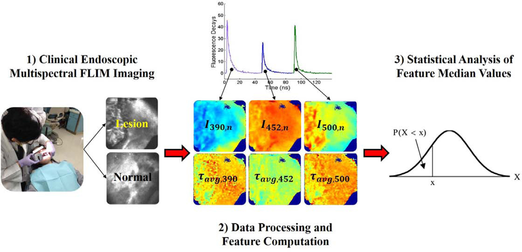
Objective: This study aimed to test the hypothesis that autofluorescence biomarkers of oral precancer and cancer can be clinically imaged and quantified by means of multispectral fluorescence lifetime imaging (FLIM) endoscopy.
Methods: Multispectral autofluorescence lifetime images of precancerous and cancerous lesions from 39 patients were imaged in vivo using a novel multispectral FLIM endoscope and processed to generate widefield maps of biochemical and metabolic autofluorescence biomarkers of oral precancer and cancer.
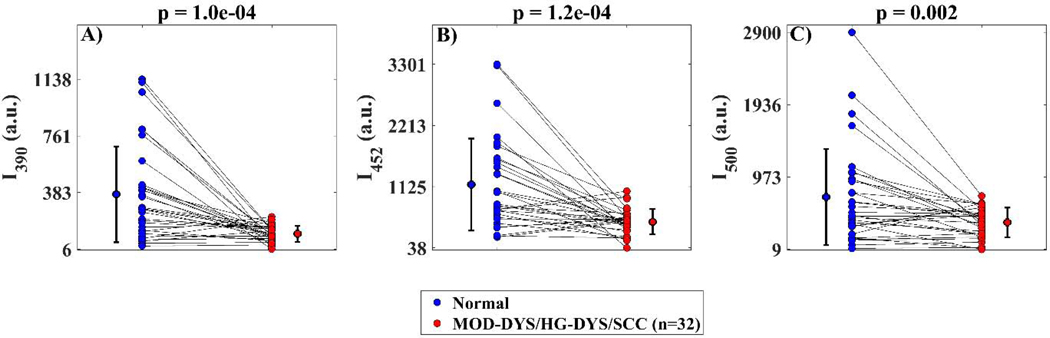
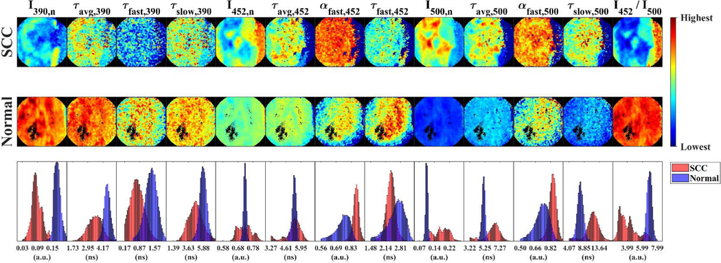
Results: Statistical analyses applied to the quantified multispectral FLIM endoscopy based autofluorescence biomarkers indicated their potential to provide contrast between precancerous/cancerous vs. healthy oral epithelial tissue.
Conclusion: To the best of our knowledge, this study represents the first demonstration of label-free biochemical and metabolic clinical imaging of precancerous and cancerous oral lesions by means of widefield multispectral autofluorescence lifetime endoscopy. Future studies will focus on demonstrating the capabilities of endogenous multispectral FLIM endoscopy as an image-guided surgical tool for positive margin detection during head and neck cancer resection surgery.
Keywords: Autofluorescence biomarkers; Fluorescence lifetime imaging (FLIM); Oral cancer and dysplasia; Statistical analysis.
Copyright © 2020 Elsevier Ltd. All rights reserved.
Conflict of interest statement
Declaration of Competing Interest The authors declare that they have no known competing financial interests or personal relationships that could have appeared to influence the work reported in this paper.
Figures

Similar articles
-
Clinical label-free endoscopic imaging of biochemical and metabolic autofluorescence biomarkers of benign, precancerous, and cancerous oral lesions.Biomed Opt Express. 2022 Jun 2;13(7):3685-3698. doi: 10.1364/BOE.460081. eCollection 2022 Jul 1. Biomed Opt Express. 2022. PMID: 35991912 Free PMC article.
-
Machine-Learning Assisted Discrimination of Precancerous and Cancerous from Healthy Oral Tissue Based on Multispectral Autofluorescence Lifetime Imaging Endoscopy.Cancers (Basel). 2021 Sep 23;13(19):4751. doi: 10.3390/cancers13194751. Cancers (Basel). 2021. PMID: 34638237 Free PMC article.
-
Computer-assisted discrimination of cancerous and pre-cancerous from benign oral lesions based on multispectral autofluorescence imaging endoscopy.Biophotonics Discov. 2024 Jul;1(2):025001. doi: 10.1117/1.bios.1.2.025001. Epub 2024 Jul 3. Biophotonics Discov. 2024. PMID: 39544621 Free PMC article.
-
Autofluorescence imaging to identify oral malignant or premalignant lesions: Systematic review and meta-analysis.Head Neck. 2020 Dec;42(12):3735-3743. doi: 10.1002/hed.26430. Epub 2020 Aug 31. Head Neck. 2020. PMID: 32866310
-
Autofluorescence Image-Guided Endoscopy in the Management of Upper Aerodigestive Tract Tumors.Int J Environ Res Public Health. 2022 Dec 22;20(1):159. doi: 10.3390/ijerph20010159. Int J Environ Res Public Health. 2022. PMID: 36612479 Free PMC article. Review.
Cited by
-
Luminescence lifetime imaging of three-dimensional biological objects.J Cell Sci. 2021 May 1;134(9):1-17. doi: 10.1242/jcs.254763. Epub 2021 May 7. J Cell Sci. 2021. PMID: 33961054 Free PMC article.
-
Perspective on the use of fluorescence molecular imaging for peripheral and deep en face margin assessment.J Biomed Opt. 2025 Jan;30(Suppl 1):S13711. doi: 10.1117/1.JBO.30.S1.S13711. Epub 2025 May 2. J Biomed Opt. 2025. PMID: 40321301 Free PMC article. Review.
-
Multidimensional quantitative characterization of the tumor microenvironment by multicontrast nonlinear microscopy.Biomed Opt Express. 2022 Sep 29;13(10):5517-5532. doi: 10.1364/BOE.470104. eCollection 2022 Oct 1. Biomed Opt Express. 2022. PMID: 36425619 Free PMC article.
-
Multimodal optical imaging with real-time projection of cancer risk and biopsy guidance maps for early oral cancer diagnosis and treatment.J Biomed Opt. 2023 Jan;28(1):016002. doi: 10.1117/1.JBO.28.1.016002. Epub 2023 Jan 13. J Biomed Opt. 2023. PMID: 36654656 Free PMC article.
-
FLIMB: fluorescence lifetime microendoscopy for metabolic and functional imaging of femoral marrow at subcellular resolution.Biomed Opt Express. 2025 Mar 31;16(4):1711-1731. doi: 10.1364/BOE.549311. eCollection 2025 Apr 1. Biomed Opt Express. 2025. PMID: 40321997 Free PMC article.
References
-
- Society AC. American Cancer Society: Cancer Facts and Figures 2019: American Cancer Society; Atlanta, GA, 2019.
-
- Kain JJ, Birkeland AC, Udayakumar N, et al. Surgical margins in oral cavity squamous cell carcinoma: Current practices and future directions. The Laryngoscope 2019 - PubMed
-
- Smits RW, Koljenović S, Hardillo JA, et al. Resection margins in oral cancer surgery: room for improvement. Head & neck 2016;38(S1):E2197–E203 - PubMed
Publication types
MeSH terms
Grants and funding
LinkOut - more resources
Full Text Sources
Medical
