Review
. 2020 Apr 4;378(3):38.
doi: 10.1007/s41061-020-0299-3.

DNA-Scaffolded Proximity Assembly and Confinement of Multienzyme Reactions

Affiliations
Review

DNA-Scaffolded Proximity Assembly and Confinement of Multienzyme Reactions

Jinglin Fu et al. Top Curr Chem (Cham). .

Abstract

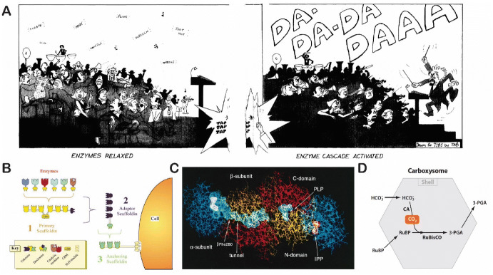
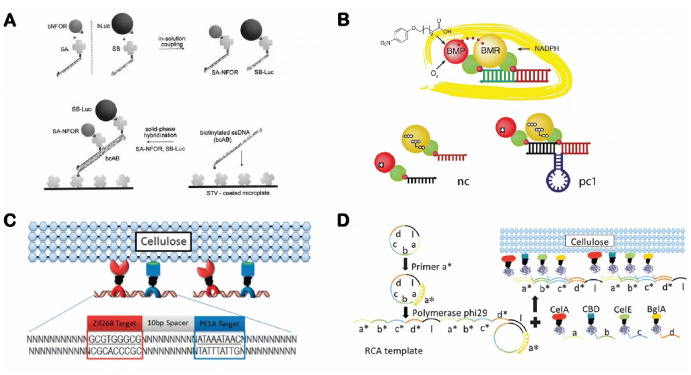
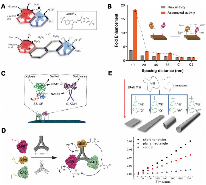
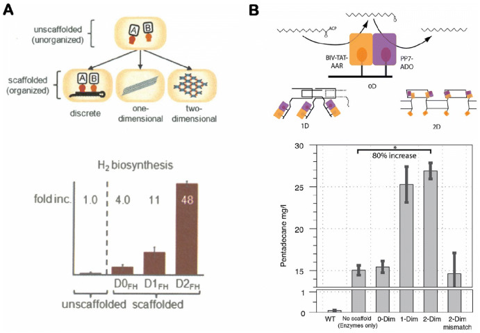
Cellular functions rely on a series of organized and regulated multienzyme cascade reactions. The catalytic efficiencies of these cascades depend on the precise spatial organization of the constituent enzymes, which is optimized to facilitate substrate transport and regulate activities. Mimicry of this organization in a non-living, artificial system would be very useful in a broad range of applications-with impacts on both the scientific community and society at large. Self-assembled DNA nanostructures are promising applications to organize biomolecular components into prescribed, multidimensional patterns. In this review, we focus on recent progress in the field of DNA-scaffolded assembly and confinement of multienzyme reactions. DNA self-assembly is exploited to build spatially organized multienzyme cascades with control over their relative distance, substrate diffusion paths, compartmentalization and activity actuation. The combination of addressable DNA assembly and multienzyme cascades can deliver breakthroughs toward the engineering of novel synthetic and biomimetic reactors.

Keywords: Biomimetic systems; DNA nanotechnology; DNA scaffolded assembly; Enzyme encapsulation; Enzyme immobilization; Enzyme regulation; Multienzyme cascade; Synthetic reactors.


Conflict of interest statement

The authors declare no competing financial interest.

Figures

Fig. 1
Fig. 1
Cellular multienzyme cascade pathways. a A cartoon of symphony performance to describe the integrated and regulatory function of enzyme cascades. Reproduced from Roach [2], with permission, copyright 1977, Trends in Biochemical Sciences. b The proximity assembly of enzymes on a protein scaffold in a cellulosome. Reproduced from Bayor et al. [5], with permission, copyright 2004, Annual Review of Microbiology. c Substrate channeling in tryptophan synthase. Reproduced from Miles et al. [6], with permission, copyright 1999, American Society for Biochemistry and Molecular Biology. d Confined carbon dioxide (CO2) fixation in a carboxysome. Reproduced from Yeates et al. [7], with permission, copyright 2010, Annual Review of Biophysics
Fig. 2
Fig. 2
Overview of structural DNA nanotechnology. a A four-way “Holliday” junction. Reproduced from Seeman [19], with permission, copyright 2003, Springer Nature. b DNA double-crossover (left) and triple-crossover (right) tiles. Reproduced from Zadegan and Norton [18], with permission, copyright 2012, MDPI. c DNA origami assembly. Reproduced from Rothemund [20], with permission, copyright 2006, Springer Nature. d Assembly of single-stranded DNA tiles. Reproduced from Wei et al. [21], with permission, copyright 2012, Springer Nature. e DNA scaffold-directed assembly of biomolecular complexes. Reproduced from Fu et al. [36], with permission, copyright 2019, John Wiley and Sons
Fig. 3
Fig. 3
Assembly of enzyme cascades on linear double-stranded DNA scaffolds. a NAD(P)H:FMN oxidoreductase and luciferase cascade. Reproduced from Niemeyer et al. [58], with permission, copyright 2002, John Wiley and Sons. b Engineered cytochrome P450 BM3 complex varying the distance between the BMR reductase domain and the BMP porphyrin domain. Reproduced from Erkelenz et al. [74], with permission, copyright 2011, American Chemical Society. c Zinc finger protein (ZFP)-appended proteins for cellulose degradation. Reproduced from Sun et al. [76], with permission, copyright 2013, Royal Society of Chemistry. d Rolling circle amplification (RCA) assembly of multienzyme nanowires to promote cellulose degradation. Reproduced from Sun and Chen [77], with permission, copyright 2016, Royal Society of Chemistry
Fig. 4
Fig. 4
Enzyme cascades organized on two-dimensional DNA nanostructures. a A glucose oxidase–horseradish peroxidase (GOx/HRP) cascade array on two-dimensional (2D) hexagonal DNA strips. Reproduced from Wilner et al. [44], with permission, copyright 2009, Springer Nature. b Organization of a GOx/HRP cascade on DNA origami tiles with controlled spacing. Reproduced from Fu et al. [45], with permission, copyright 2012, American Chemical Society. c Assembly of an NAD cofactor-coupled enzyme cascade (XR xylose reductase, XDH xylitol dehydrogenase). Reproduced from Ngo et al. [81], with permission, copyright 2016, American Chemical Society. d A three-enzyme (MDH malic dehydrogenase, OAD oxaloacetate decarboxylase, LDH lactate dehydrogenase) cascade organized on a triangular DNA origami structure. Reproduced from Liu et al. [83], with permission, copyright 2016, John Wiley and Sons. e A rectangular DNA origami rolling into a DNA nanotube for assembly of an enzyme cascade. Reproduced from Fu et al. [80], with permission, copyright 2013, American Chemical Society
Fig. 5
Fig. 5
In vivo assembly of enzyme cascades on RNA nanostructures. a Organization of [FeFe]-hydrogenase and ferredoxin on one-dimensional (1D) and 2D RNA nanostructures (top) with enhanced hydrogen production in vivo (bottom). Reproduced from Delebecque et al. [85], with permission, copyright 2011, The American Association for the Advancement of Science. b Assembly of a two-enzyme pentadecane production pathway on RNA scaffolds (top) with enhanced pentadecane output in vivo (bottom). Reproduced from Sachdeva et al. [86], with permission, copyright 2014, Oxford University Press
Fig. 6
Fig. 6
Theoretical modeling of distance-dependent enzyme cascade reactions. a Brownian diffusion of H2O2 in a GOx/HRP reaction depending on distance [n(r, t) Number of molecules at a distance r from the initial produced position of production]. Reproduced from Fu et al. [45], with permission, copyright 2012, American Chemical Society. b Concentration profiles of the reaction product as a function of radial distance (r) from an active site under different ratios of turnover frequency to diffusion coefficient (k/D). Reproduced from Wheeldon et al. [90], with permission, copyright 2016, Springer Nature
Fig. 7
Fig. 7
Development of DNA nanocages for enzyme encapsulation. a The first 3D DNA cube. Reproduced from Seeman [19], with permission, copyright 2003, Springer Nature. b A DNA tetrahedron for encapsulating a protein. Reproduced from Erben et al. [107], with permission, copyright 2006, John Wiley and Sons. c DNA polyhedrons for organizing proteins (STV streptavidin). Reproduced from Zhang et al. [109], with permission, copyright 2019, John Wiley and Sons. d The combination of half DNA nanocages for enclosing enzymes. Reproduced from Zhao et al. [94], with permission, copyright 2016, Springer Nature. e Protein encapsulation into a DNA host using noncovalent protein–ligand interactions. Reproduced from Sprengel et al. [72] under the terms and conditions of the Creative Commons Attribution 4.0 International License, copyright 2017, Springer Nature
Fig. 8
Fig. 8
DNA nanocage-regulated encapsulation and release of protein cargoes. a Single-step folding of DNA nanotubes for enzyme encapsulation. Reproduced from Fu et al. [80], with permission, copyright 2013, American Chemical Society. b A temperature-sensitive DNA nanocage for encapsulating and releasing an enzyme. Reproduced from Juul et al. [111], with permission, copyright 2013, American Chemical Society. c A light-triggered release of bioactive cargoes from a DNA nanocage. Reproduced from Kohman et al. [112], with permission, copyright 2016, American Chemical Society. d A pH-switchable DNA tetrahedron for regulating protein stability and activity. Reproduced from Kim et al. [113], with permission, copyright 2017, American Chemical Society. e A DNA nanovault with reversible opening and closing to regulate enzyme–substrate accessibility. Reproduced from Grossi et al. [114] under the terms and conditions of the Creative Commons Attribution 4.0 International License, copyright 2017, Springer Nature
Fig. 9
Fig. 9
Enhancement of enzyme activity by DNA nanostructures. Reproduced from Zhang and Hess [115], with permission, copyright 2017, American Chemical Society
Fig. 10
Fig. 10
Biomimetic assembly of swinging arms. a Swinging domains in pyruvate dehydrogenase complex. Reproduced from Perham [121], with permission, copyright 2000, Annual Review of Biochemistry. b An artificial swinging arm to transfer NAD+ cofactor between two dehydrogenases (G6PDH glucose-6-phosphate dehydrogenase and MDH) on DNA nanoscaffolds. c Enhanced enzyme cascade activity by swinging arms. d Improved reaction selectivity by swinging arms. Reproduced from Fu et al. [47], with permission, copyright 2014, Springer Nature
Fig. 11
Fig. 11
Large biomolecular nanostructures organized by artificial swinging arms. a NAD+ arms for regulating pathway activity between the G6PDH–MDH cascade and the G6PDH–LDH cascade. Reproduced from Ke et al. [123], with permission, copyright 2016, John Wiley and Sons. b 2D enzyme arrays of the G6PDH–MDH reaction with NAD+ swinging arms. Reproduced from Yang et al. [125], with permission, copyright 2018, John Wiley and Sons
Fig. 12
Fig. 12
DNA swinging arms for facilitating bioelectroactive reactions. a A GOx–ferrocene–electrode contact. Reproduced from Piperberg et al. [126], with permission, copyright 2009, American Chemical Society. b A nitrite reductase–pseudoazurin (NiR-Paz) system (SAM self-assembled monolayer). Reproduced from Tepper [127], with permission, copyright 2010, American Chemical Society. c A TiO2–CdS complex for H2 production. Reproduced from Ma et al. [128], with permission, copyright 2015, John Wiley and Sons
Fig. 13
Fig. 13
DNA arm-based nanorobotic system. a Double-stranded DNA (dsDNA) arms for fluorescent cargo transport. Reproduced from Kopperger et al. [129], with permission, copyright 2015, American Chemical Society. b A nanoscale robotic arm driven by an electric field. Reproduced from Kopperger et al. [130], with permission, copyright 2018, The American Association for the Advancement of Science. c DNA origami rotor driven by a motor protein (left), atomic force microscopy images of a DNA rotor (middle) and origami-rotor-based imaging and tracking (ORBIT) for tracking DNA rotation (right). Reproduced from Kosuri et al. [131], with permission, copyright 2019, Springer Nature
Fig. 14
Fig. 14
Synthetic scramblase built from DNA. a The structure of a DNA-based scramblase with two cholesterol prosthetic groups. b Molecular dynamic simulation of the lipid scrambling process. Reproduced from Ohmann et al. [132] under the terms and conditions of the Creative Commons Attribution 4.0 International License, copyright 2018, Springer Nature
Fig. 15
Fig. 15
Artificial photosynthetic systems organized by DNA nanoscaffolds. a Multicolor fluorophore array for photo-energy transfer. Reproduced from Stein et al. [133], with permission, copyright 2011, American Chemical Society. b Artificial light-harvesting network. Reproduced from Dutta et al. [134], with permission, copyright 2011, American Chemical Society. c A DNA-directed light-harvesting/reaction center. Reproduced from Dutta et al. [135], with permission, copyright 2014, American Chemical Society. d A synthetic DNA-based excitonic circuit based on J-aggregates (PIC pseudoisocyanine). Reproduced from Boulais et al. [136], with permission, copyright 2017, Springer Nature
Fig. 16
Fig. 16
DNA hybridization-regulated enzyme activity. a An enzyme–inhibitor interaction (ssDNA Single-stranded DNA). Reproduced from Saghatelian et al. [138], with permission, copyright 2003, American Chemical Society. b Streptokinase–plasminogen (SK–Pg) complex for regulating fibrinolytic activity. Reproduced from Mukherjee et al. [140], with permission, copyright 2018, American Chemical Society. c Enzyme actuation (E enzyme, I inhibitor). Reproduced from Janssen [141], with permission, copyright 2015, American Chemical Society. d Photo-regulated thrombin-aptamer complex. Reproduced from Kim et al. [142], with permission, copyright 2009, National Academy of Sciences
Fig. 17
Fig. 17
DNA nanotweezers-regulated enzyme reaction. a A G6PDH/NAD+ pair. Reproduced from Liu et al. [48], with permission. Copyright 2013, Springer Nature. b A GOx/HRP cascade. Reproduced from Xin et al. [143], with permission, copyright 2013, John Wiley and Sons. c A three-component biocatalytic cascade of β-Gal/GOx/hemin. Reproduced from Hu et al. [144], with permission, copyright 2014, John Wiley and Sons. d Distance regulation of a GOx/HRP pair. Reproduced from Kou et al. [145], with permission, copyright 2018, American Chemical Society. e A trident-shaped DNA nanomachine with several conformational states for regulating an enzyme cascade. Reproduced from Xing et al. [146], with permission, copyright 2018, American Chemical Society
Fig. 18
Fig. 18
DNA hairpin-mediated proximity assembly of an enzyme and a cofactor. a Enzyme activities for a hairpin-locked cofactor, opened cofactor and co-assembled enzyme/cofactor pair. b A design chart of a biochemical sensing circuit. c Detection of microRNA and adenosine. Reproduced from Oh et al. [49], with permission, copyright 2018, John Wiley and Sons

Similar articles

Cited by

References

    1. Schomburg I, Chang A, Placzek S, Söhngen C, Rother M, Lang M, Munaretto C, Ulas S, Stelzer M, Grote A, Scheer M, Schomburg D. BRENDA in 2013: integrated reactions, kinetic data, enzyme function data, improved disease classification: new options and contents in BRENDA. Nucleic Acids Res. 2013;41:D764–D772. - PMC - PubMed
    1. Roach PJ. Functional significance of enzyme cascade systems. Trends Biochem Sci. 1977;2:87–90.
    1. Savage DF, Afonso B, Chen AH, Silver PA. Spatially ordered dynamics of the bacterial carbon fixation machinery. Science. 2010;327:1258–1261. - PubMed
    1. Srere PA, Mosbach K. Metabolic compartmentation: symbiotic, organellar, multienzymic, and microenvironmental. Annu Rev Microbiol. 1974;28:61–84. - PubMed
    1. Bayer EA, Belaich JP, Shoham Y, Lamed R. The cellulosomes: multienzyme machines for degradation of plant cell wall polysaccharides. Annu Rev Microbiol. 2004;58:521–554. - PubMed

LinkOut - more resources