

Metformin Improves Cardiac Metabolism and Function, and Prevents Left Ventricular Hypertrophy in Spontaneously Hypertensive Rats
- PMID: 32248762
- PMCID: PMC7428616
- DOI: 10.1161/JAHA.119.015154
Metformin Improves Cardiac Metabolism and Function, and Prevents Left Ventricular Hypertrophy in Spontaneously Hypertensive Rats
Erratum in
-
Correction to: Metformin Improves Cardiac Metabolism and Function, and Prevents Left Ventricular Hypertrophy in Spontaneously Hypertensive Rats.J Am Heart Assoc. 2020 Jun 16;9(12):e014528. doi: 10.1161/JAHA.119.014528. Epub 2020 May 23. J Am Heart Assoc. 2020. PMID: 32448022 Free PMC article. No abstract available.
Abstract
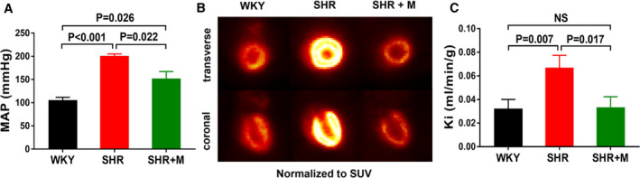
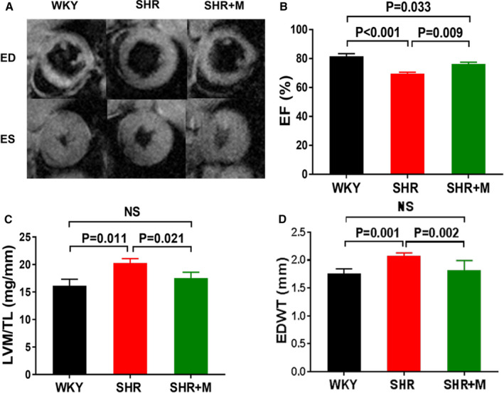
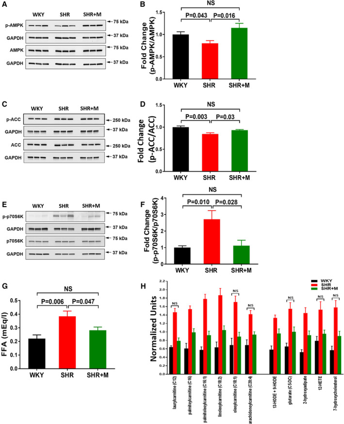
Background In spontaneously hypertensive rats (SHR) we observed profound myocardial metabolic changes during early hypertension before development of cardiac dysfunction and left ventricular hypertrophy. In this study, we evaluated whether metformin improved myocardial metabolic abnormalities and simultaneously prevented contractile dysfunction and left ventricular hypertrophy in SHR. Methods and Results SHR and control Wistar-Kyoto rats were treated with metformin from 2 to 5 months of age, when SHR hearts exhibit metabolic abnormalities and develop cardiac dysfunction and left ventricular hypertrophy. We evaluated the effect of metformin on myocardial glucose uptake rates with dynamic 2-[18F] fluoro-2-deoxy-D-glucose positron emission tomography. We used cardiac MRI in vivo to assess the effect of metformin on ejection fraction, left ventricular mass, and end-diastolic wall thickness, and also analyzed metabolites, AMP-activated protein kinase and mammalian target-of-rapamycin activities, and mean arterial blood pressure. Metformin-treated SHR had lower mean arterial blood pressure but remained hypertensive. Cardiac glucose uptake rates, left ventricular mass/tibia length, wall thickness, and circulating free fatty acid levels decreased to normal, and ejection fraction improved in treated SHR. Hearts of treated SHR exhibited increased AMP-activated protein kinase phosphorylation and reduced mammalian target-of-rapamycin activity. Cardiac metabolite profiling demonstrated that metformin decreased fatty acyl carnitines and markers of oxidative stress in SHR. Conclusions Metformin reduced blood pressure, normalized myocardial glucose uptake, prevented left ventricular hypertrophy, and improved cardiac function in SHR. Metformin may exert its effects by normalizing myocardial AMPK and mammalian target-of-rapamycin activities, improving fatty acid oxidation, and reducing oxidative stress. Thus, metformin may be a new treatment to prevent or ameliorate chronic hypertension-induced left ventricular hypertrophy.
Keywords: cardiac MRI; cardiac hypertrophy; dynamic FDG PET; metformin; spontaneously hypertensive rats.
Figures
References
-
- Cuspidi C, Facchetti R, Bombelli M, Tadic M, Sala C, Grassi G, Mancia G. High normal blood pressure and left ventricular hypertrophy echocardiographic findings from the PAMELA population. Hypertension. 2019;73:612–619. - PubMed
-
- Levy D, Garrison RJ, Savage DD, Kannel WB, Castelli WP. Prognostic implications of echocardiographically determined left ventricular mass in the Framingham Heart Study. N Engl J Med. 1990;322:1561–1566. - PubMed
-
- Verdecchia P, Carini G, Circo A, Dovellini E, Giovannini E, Lombardo M, Solinas P, Gorini M, Maggioni AP. Left ventricular mass and cardiovascular morbidity in essential hypertension: The MAVI study. J Am Coll Cardiol. 2001;38:1829–1835. - PubMed
-
- Devereux RB, Wachtell K, Gerdts E, Boman K, Nieminen MS, Papademetriou V, Rokkedal J, Harris K, Aurup P, Dahlof B. Prognostic significance of left ventricular mass change during treatment of hypertension. JAMA. 2004;292:2350–2356. - PubMed
-
- Li J, Kemp BA, Howell NL, Massey J, Minczuk K, Huang Q, Chordia MD, Roy RJ, Patrie JT, Davogustto GE, et al. Metabolic changes in spontaneously hypertensive rat hearts precede cardiac dysfunction and left ventricular hypertrophy. J Am Heart Assoc. 2019;8:e010926 DOI: 10.1161/JAHA.118.010926. - DOI - PMC - PubMed
Publication types
MeSH terms
Substances
Grants and funding
LinkOut - more resources
Full Text Sources
Medical
