Review
. 2020 May 25;375(1799):20190230.
doi: 10.1098/rstb.2019.0230. Epub 2020 Apr 6.

A mechanism for learning with sleep spindles

Affiliations
Review

A mechanism for learning with sleep spindles

Adrien Peyrache et al. Philos Trans R Soc Lond B Biol Sci. .

Abstract

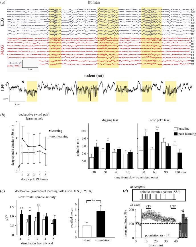
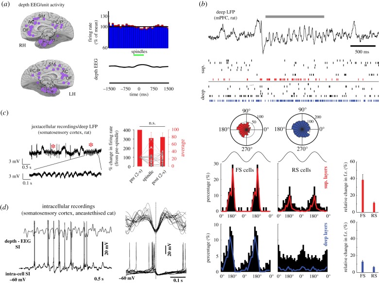
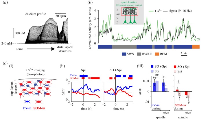
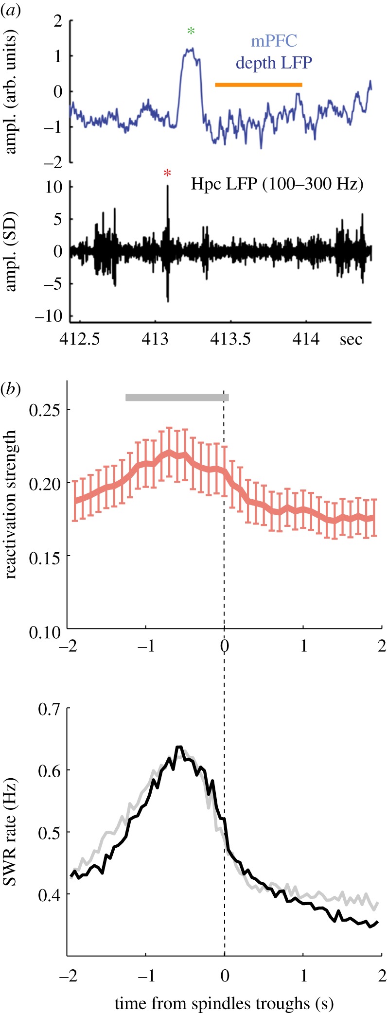
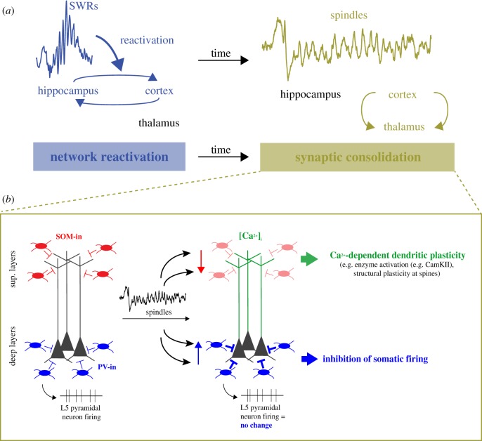
Spindles are ubiquitous oscillations during non-rapid eye movement (NREM) sleep. A growing body of evidence points to a possible link with learning and memory, and the underlying mechanisms are now starting to be unveiled. Specifically, spindles are associated with increased dendritic activity and high intracellular calcium levels, a situation favourable to plasticity, as well as with control of spiking output by feed-forward inhibition. During spindles, thalamocortical networks become unresponsive to inputs, thus potentially preventing interference between memory-related internal information processing and extrinsic signals. At the system level, spindles are co-modulated with other major NREM oscillations, including hippocampal sharp wave-ripples (SWRs) and neocortical slow waves, both previously shown to be associated with learning and memory. The sequential occurrence of reactivation at the time of SWRs followed by neuronal plasticity-promoting spindles is a possible mechanism to explain NREM sleep-dependent consolidation of memories. This article is part of the Theo Murphy meeting issue 'Memory reactivation: replaying events past, present and future'.

Keywords: coupling; memory; plasticity; reactivation; sleep; spindles.


Conflict of interest statement

We have no competing interests.

Figures

Figure 1.
Figure 1.
Spindles, memory and plasticity. (a) Examples of spindles in human electroencephalograph (EEG) and magnetoencephalogram (MAG) (upper traces, adapted from [37]) and rat (lower trace; J. Seibt 2017, unpublished data). Spindles are highlighted in yellow. (b) Influence of learning on spindle density in the human (left graph, adapted from [38]) and rat (right graphs, adapted from [39]) frontal cortex. (c) Boosting frontal slow spindles (and SOs, not shown) with frontolateral slow oscillatory (0.75 Hz) transcranial direct current stimulation (so-tDCS) in humans improves declarative memory (adapted from [21]). (d) Naturalistic spindle stimulus pattern (SSP) promotes short- (STP) and long-term plasticity (LTP) in vitro (adapted from [40]).
Figure 2.
Figure 2.
Inhibition of cortical activity during spindles. (a) Depth EEG/unit activity recordings in various brain regions (purple circles) in humans. Average cortical firing rate (across various depths and brain regions) during spindles (green bar) (adapted from [43]). (b) Entrainment of regular spiking (RS, putative excitatory) and fast spiking (FS, putative inhibitory) neurons during spindles in the superficial and deep layers of rat medial prefrontal cortex (mPFC). Top trace and raster: example of a spindle event (grey bar) and associated firing in the superficial and deep layers of the mPFC. The red and blue dots in the raster indicate spike times of two putative FS neurons in the superficial (sup.) and deep layers, respectively. Polar histograms show the distribution of the phases of these two neurons relative to all spindles. Bottom left: distribution of preferred phases to spindles for all neurons across layers and cell types (black histogram) and only for the significantly modulated neurons (colour histograms). Bottom right: changes in firing rate (f.r.) during spindles relative to NREM sleep across cell types and layers (adapted from [92]). (c) Average change in firing rate peri-spindles (red stars) of L5 pyramidal neurons in the somatosensory cortex of head-fixed rats (adapted from [31]). (d) Intracellular recordings of pyramidal neurons activity during spindles in the primary somatosensory cortex (SI) of anaesthetized cats. Left: example of spindles sequence recorded with depth electroencephalogram (depth - EEG) together with neuronal discharge. Right: superimposition of 10 spindle cycles of the same cell. Traces were aligned to negative peaks of depth EEG. Note the excitatory post-synaptic potential followed by inhibitory post-synaptic potential followed by IPSP during spindles (adapted from [93]).
Figure 3.
Figure 3.
Cortical microcircuit regulation during spindle activity. (a) Model of predicted deep layers (L5/6) somatic and dendritic intracellular Ca2+ activity during spindles. Instantaneous profiles taken every 0.2 ms are superimposed. Note the high Ca2+ activity only in dendrites and not in soma (adapted from [93]). (b) In vivo Ca2+ imaging (photometry) in apical dendrites of L5 neurons (somatosensory cortex) in freely behaving and sleeping rats. Superimposed activity of population of dendrites and sigma power for an approximately 30 min recording in a freely moving rat (adapted from [31]). (c) (i) In vivo Ca2+ imaging (two-photon) of cortical inhibitory neuron subpopulations (PV-in: parvalbumin inhibitory; SOM-in: somatostatin inhibitory) in superficial layers of the cortex. (ii) Mean Ca2+ activity aligned to spindles with or without SO coupling. Bars indicate significant differences compared to baseline. (iii) Ca2+ activity changes during and after spindle occurrence (adapted from [101]).
Figure 4.
Figure 4.
SWR and spindles comodulation and link to replay. (a) Example traces of LFP recorded simultaneously in the medial prefrontal cortex (mPFC, top, broadband) and in the CA1 pyramidal layer of the hippocampus (Hpc, bottom, filtered in the SWR-frequency range) (adapted from Peyrache et al. [92]). Red and green asterisks indicate a SWR and a SW, respectively. Spindles are highlighted with an orange bar. (b) Relationship between spindles and reactivation in the mPFC. Top: spindle-trough time average (± s.e.m.) of reactivation strength of neuronal ensembles during sleep following learning; bottom, same as top for SWR occurrence rate (adapted from [111]).
Figure 5.
Figure 5.
The sequential hypothesis of hippocampo-cortical coupling and plasticity role of spindles. (a) Illustration of the sequential hypothesis of hippocampo-cortical coupling. Reactivation and hippocampo-cortical network interactions during SWRs are followed by spindle events during which synaptic communication in those networks are modified via synaptic plasticity mechanisms. (b) Model of cortical circuit activity regulation of deep layers pyramidal neurons during spindles based on Ca2+ imaging, electrophysiological and computational data. During spindles, dendrites are disinhibited via decreased activity of somatostatin inhibitory (SOM-In) neurons while somatic spiking is inhibited via increase activity of parvalbumin inhibitory (PV-In) neurons. This would lead to Ca2+-dependent plasticity processes (e.g. CaMKII activation) occurring specifically in dendrites.

Similar articles

Cited by

References

    1. Berger H. 1929. Über das Elektrenkephalogramm des Menschen. Arch. Für Psychiatr. Nervenkrankh. 87, 527–570. (10.1007/BF01797193) - DOI
    1. Loomis AL, Harvey EN, Hobart G. 1935. Potential rhythms of the cerebral cortex during sleep. Science 81, 597–598. (10.1126/science.81.2111.597) - DOI - PubMed
    1. Coppieters ‘t Wallant D, Maquet P, Phillips C. 2016. Sleep spindles as an electrographic element: description and automatic detection methods. Neural Plast. 2016, 6783812 (10.1155/2016/6783812) - DOI - PMC - PubMed
    1. De Gennaro L, Ferrara M. 2003. Sleep spindles: an overview. Sleep Med. Rev. 7, 423–440 (10.1053/smrv.2002.0252). - DOI - PubMed
    1. Fernandez LMJ, Luthi A. 2019. Sleep spindles: mechanisms and functions. Physiol. Rev. 100, 805–868. (10.1152/physrev.00042.2018) - DOI - PubMed

Publication types

LinkOut - more resources