. 2020 May:55:102743.
doi: 10.1016/j.ebiom.2020.102743. Epub 2020 Apr 2.

Microneedle array delivered recombinant coronavirus vaccines: Immunogenicity and rapid translational development

Affiliations

Microneedle array delivered recombinant coronavirus vaccines: Immunogenicity and rapid translational development

Eun Kim et al. EBioMedicine. 2020 May.

Abstract

Background: Coronaviruses pose a serious threat to global health as evidenced by Severe Acute Respiratory Syndrome (SARS), Middle East Respiratory Syndrome (MERS), and COVID-19. SARS Coronavirus (SARS-CoV), MERS Coronavirus (MERS-CoV), and the novel coronavirus, previously dubbed 2019-nCoV, and now officially named SARS-CoV-2, are the causative agents of the SARS, MERS, and COVID-19 disease outbreaks, respectively. Safe vaccines that rapidly induce potent and long-lasting virus-specific immune responses against these infectious agents are urgently needed. The coronavirus spike (S) protein, a characteristic structural component of the viral envelope, is considered a key target for vaccines for the prevention of coronavirus infection.

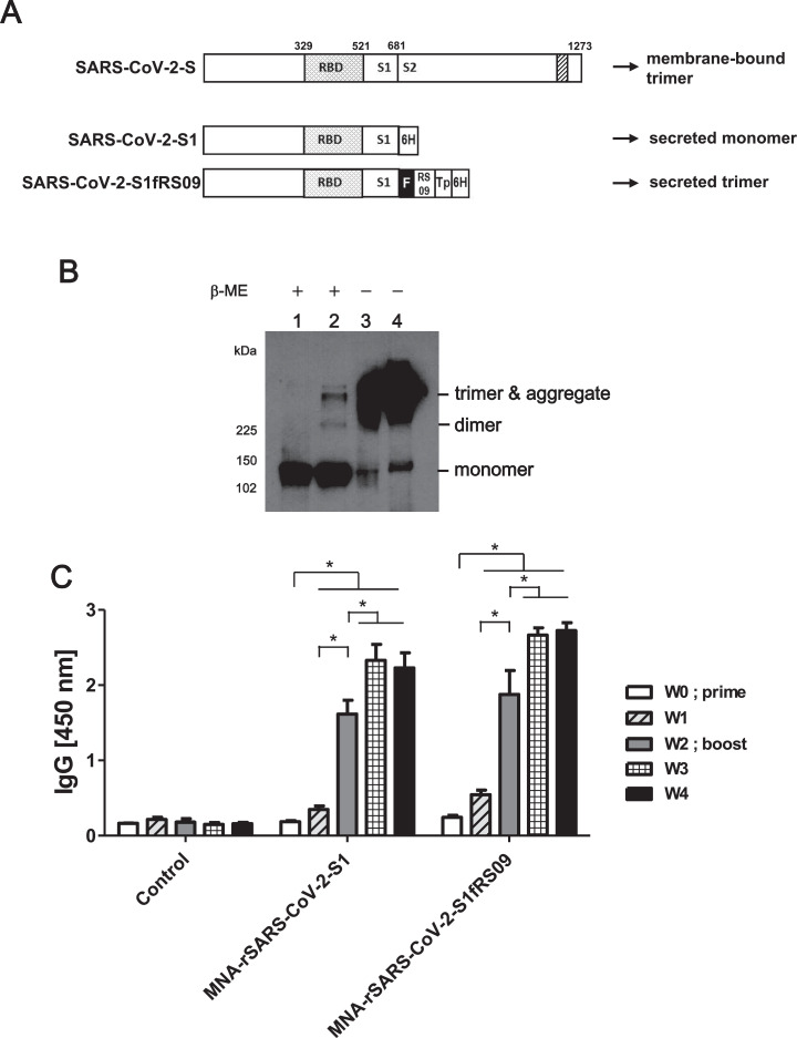
Methods: We first generated codon optimized MERS-S1 subunit vaccines fused with a foldon trimerization domain to mimic the native viral structure. In variant constructs, we engineered immune stimulants (RS09 or flagellin, as TLR4 or TLR5 agonists, respectively) into this trimeric design. We comprehensively tested the pre-clinical immunogenicity of MERS-CoV vaccines in mice when delivered subcutaneously by traditional needle injection, or intracutaneously by dissolving microneedle arrays (MNAs) by evaluating virus specific IgG antibodies in the serum of vaccinated mice by ELISA and using virus neutralization assays. Driven by the urgent need for COVID-19 vaccines, we utilized this strategy to rapidly develop MNA SARS-CoV-2 subunit vaccines and tested their pre-clinical immunogenicity in vivo by exploiting our substantial experience with MNA MERS-CoV vaccines.

Findings: Here we describe the development of MNA delivered MERS-CoV vaccines and their pre-clinical immunogenicity. Specifically, MNA delivered MERS-S1 subunit vaccines elicited strong and long-lasting antigen-specific antibody responses. Building on our ongoing efforts to develop MERS-CoV vaccines, promising immunogenicity of MNA-delivered MERS-CoV vaccines, and our experience with MNA fabrication and delivery, including clinical trials, we rapidly designed and produced clinically-translatable MNA SARS-CoV-2 subunit vaccines within 4 weeks of the identification of the SARS-CoV-2 S1 sequence. Most importantly, these MNA delivered SARS-CoV-2 S1 subunit vaccines elicited potent antigen-specific antibody responses that were evident beginning 2 weeks after immunization.

Interpretation: MNA delivery of coronaviruses-S1 subunit vaccines is a promising immunization strategy against coronavirus infection. Progressive scientific and technological efforts enable quicker responses to emerging pandemics. Our ongoing efforts to develop MNA-MERS-S1 subunit vaccines enabled us to rapidly design and produce MNA SARS-CoV-2 subunit vaccines capable of inducing potent virus-specific antibody responses. Collectively, our results support the clinical development of MNA delivered recombinant protein subunit vaccines against SARS, MERS, COVID-19, and other emerging infectious diseases.

Keywords: COVID-19; MERS-CoV S1; Microneedle array; SARS-CoV-2; Subunit vaccines; Trimerization.


Figures

Fig 1
Fig. 1
Construction of recombinant MERS-S1 Foldon subunit vaccines. (A) Schematic diagram of rMERS-S1s. The positions of the RBD (small dots) and transmembrane domain (stripes) are indicated and S is divided into two subdomains, S1 and S2, at position 751. The vector was used to generate recombinant replication-deficient adenoviruses by homologous recombination with the adenoviral genomic DNA. Arrows showed the cleavage site by TEV protease. Abbreviations are as follows: ITR, inverted terminal repeat; RBD, receptor binding domain;F, T4 fibritin foldon trimerization domain; Tp, Tobacco Etch Virus (TEV) protease; fliC; Salmonella typhimurium flagellin C (B) Western blot of the supernatant of A549 cells infected (10 MOI) with mock (lane 1 and 5), Ad5.MERS-S1f (lane 2 and 6), Ad5.MERS-S1fRS09 (lane 3 and 7), or Ad5.MERS-S1ffliC (lane 4 and 8), respectively, with anti-6His monoclonal antibody. The supernatants were resolved on SDS-4~20% polyacrylamide gel after being boiled in 2% SDS sample buffer with or without β-ME. (C) Detection of purified rMERS-S1fs with mouse sera against Ad5.MERS-S1. The recombinant MERS-S1f proteins were purified using His60 Ni Superflow Resin under native conditions. After cleavage of the fusion protein by TEV protease, 6xHis tag and protease were removed from the cleavage reaction by affinity chromatography on a nickel chelating resin. Three rMERS-S1fs were coated in 96-well plate with 200ng/well ELISA and detected with mouse sera against Ad5.MERS-S1. Significance was determined by one-way ANOVA followed by Tukey's test. *p < 0.05.
Fig 2
Fig. 2
MNA delivered subunit vaccines elicit antibodies recognizing cell surface bound MERS-S proteins. (A) The experimental schedule representing the timeline for the immunizations. (B) Flow cytometry assay of 293 cells expressing MERS-S at the cell surface. HEK 293 cells were transfected with pAd/MERS-S (gray bar) or control pAd (white bar). At 36 h post-transfection, binding to MERS-S at the cell surface was analyzed by incubation with mice sera obtained at week 2 after immunization followed by staining with PE-conjugated anti-mouse IgG. St Significance was determined by one-way ANOVA followed by Tukey's test. *p < 0.05.
Fig 3
Fig. 3
Induction of humoral immune response in mice vaccinated with recombinant MERS-S1 vaccines. BALB/c mice were immunized subcutaneously or intracutaneously with 20 µg of each rMERS-S1 subunit vaccines s.c., with or without 20 µg of MPLA, or by MNA delivery of the same subunit proteins, or with 1011vp of Ad5.MERS-S1 as a positive control. On day 14 mice were boosted using the same regimen as the prime immunization. On weeks 0, 2, 4, and 6 after treatment, immune sera from mice were collected, diluted (200x), and tested for the presence of MERS-S1-specific antibodies by ELISA (A and B) or by MERS-CoV-neutralization assay without dilution (C and D). MERS-CoV virus-neutralizing titers (VNTs) were measured every week after primary immunization using Vero cells by determining the highest dilution inhibiting MERS-CoV infection by 100%. Significance was determined by one-way ANOVA followed by Tukey's test. *p < 0.05.White and black circles in Fig. 3C and D represent week 4 and week 6, respectively.
Fig 4
Fig. 4
Longevity of immune response in mice vaccinated with subunit vaccines. BALB/c mice were immunized with subunit vaccines as described in the Fig. 3 legend. On week 23 and 55 after immunization sera were collected, diluted (200x) in PBS, and tested for the presence of MERS-S1-specific antibodies by ELISA. Significance was determined by one-way ANOVA followed by Tukey's test. *p < 0.05.
Fig 5
Fig. 5
Induction of humoral immune response in mice immunized with MNA delivered SARS-CoV-2-S1 vaccines. (A) Schematic diagram of rSARS-CoV-2-S1s. The positions of the RBD (small dots) and transmembrane domain (stripes) are indicated and S is divided into two subdomains, S1 and S2, at position 681. The SARS-CoV-2-S1 or SARS-CoV-2-S1fRS09 gene was inserted into pmax Cloning expression vector. (B) Western blot of the purified rSARS-CoV-2-S1 (lane 1 and 3) or rSARS-CoV-2-S1fRS09 (lane 2 and 4) proteins with anti-6His monoclonal antibody. The purified proteins were resolved on 10% Tris/Glycine gel after being boiled in 2% SDS sample buffer with β-ME or in native sample buffer without β-ME. (C) C57BL/6 mice were immunized intradermally with MNAs containing 20 µg of rSARS-CoV-2-S1 or rSARS-CoV-2-S1fRS09. Immune sera from mice were collected every week, diluted (100x) in PBS, and tested for the presence of SARS-CoV-2-S1-specific antibodies by ELISA. Significance was determined by one-way ANOVA followed by Tukey's test. *p < 0.05.

References

    1. Zaki AM, van Boheemen S, Bestebroer TM, Osterhaus AD, Fouchier RA. Isolation of a novel coronavirus from a man with pneumonia in Saudi Arabia. N Engl J Med. 2012;367(19):1814–1820. - PubMed
    1. (WHO) WHO. Middle East respiratory syndrome coronavirus (MERS-CoV). Geneva, Switzerland. Available from: http://www.who.int/emergencies/mers-cov/en/. 2018[cited 2018 November 20].
    1. Nishiura H, Endo A, Saitoh M. Identifying determinants of heterogeneous transmission dynamics of the Middle East respiratory syndrome (MERS) outbreak in the Republic of Korea, 2015: a retrospective epidemiological analysis. BMJ Open. 2016;6(2) - PMC - PubMed
    1. Park HY, Lee EJ, Ryu YW. Epidemiological investigation of MERS-CoV spread in a single hospital in South Korea, May to June 2015. Euro Surveill Bull Eur Sur Les Malad Transm Eur Commun Disease Bull. 2015;20(25):1–6. - PubMed
    1. Al-Omari A, Rabaan AA, Salih S, Al-Tawfiq JA, Memish ZA. MERS coronavirus outbreak: Implications for emerging viral infections. Diagnostic Microbiol Infect Disease. 2018 - PMC - PubMed

MeSH terms