Review
. 2020 Jun:33:101516.
doi: 10.1016/j.redox.2020.101516. Epub 2020 Mar 23.

Endoplasmic reticulum stress and glutathione therapeutics in chronic lung diseases

Affiliations
Review

Endoplasmic reticulum stress and glutathione therapeutics in chronic lung diseases

Yvonne Janssen-Heininger et al. Redox Biol. 2020 Jun.
No abstract available

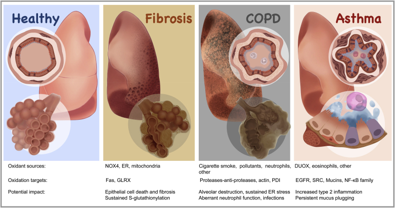
Keywords: Asthma; Chronic obstructive pulmonary disease; Endoplasmic reticulum; Fibrosis; Glutaredoxin; Glutathione S-transferase P; Idiopathic pulmonary fibrosis; Mitochondria; Protein disulfide isomerase; S-glutathionylation; Unfolded protein response.


Conflict of interest statement

Declaration of competing interest Yvonne Janssen-Heininger, Niki L. Reynaert, and Vikas Anathy hold patents: United States Patent No. 8,679,811, “Treatments Involving Glutaredoxins and Similar Agents” (YJ-H, VA), United States Patent No. 8,877,447, “Detection of Glutathionylated Proteins” (YJ-H, NLR), United States Patent, 9,907,828, “Treatments of oxidative stress conditions” (YJ-H, VA) Yvonne Janssen-Heininger and Vikas Anathy have received consulting fees from Celdara Medical LLC for their contributions with the commercialization of glutaredoxin for the treatment of pulmonary fibrosis.

Figures

Fig. 1
Fig. 1
Steps in the catalytic cycle of protein S-glutathionylation (PSSG) and deglutathionylation believed to be relevant in the pathogenesis of chronic lung diseases. A number of biochemical events can induce PSSG. The mode of PSSG is likely to be target and context specific, dependent upon the proximity of oxidant producing enzymes, redox relays, the oxidation state of the GSH/GSSG redox couple etc. In epithelial cells in settings of a pro-fibrotic environment, oxidants originating from multiple sources lead to a sulfenic acid intermediate (SOH) which can be a platform for subsequent PSSG, which can happen spontaneously or catalyzed by glutathione S- transferases (GST), notably GSTP [1]. Conversely GLRX acts to deglutathionylate proteins [2], restoring the original sulfhydryl group. GLRX induces deglutathionylation via the monothiol mechanism requiring only the N-terminal cysteine in the thioredoxin (TXN) domain [2], which can thereafter be reduced by reduced glutathione (GSH) [3] re-establishing the reduced thiol group of the N-terminal cysteine in GLRX. Glutathione disulfide (GSSG), formed in this process can be reduced back to GSH through the action of glutathione reductase (GR) consuming reducing equivalents form NADPH [4]. GLRX can be inactivated via oxidations of one or more cysteines outside of the TXN domain [5] including electrophiles from cigarette smoke. GLRX can also directly cleaved by caspases 8 and 3 [6]. Similarly, GSTP also is subject to oxidant-mediated inactivation, potentially involving a disulfide which creates steric hindrance that interferes with GSH binding [7], or electrophile-induced modification [8]. We refer the reader to the body of text for detailed information. Red circle: protein cysteine or cysteine in glutathione. SH: reduced cysteine, SOH: Sulfenic acid, SSG: S-glutathionylated protein, GSH: Reduced glutathione, GSSG: glutathione disulfide, comprised of two glutathione molecules with a disulfide bond between the cysteines of each glutathione molecule, S–S: disulfide. (For interpretation of the references to colour in this figure legend, the reader is referred to the Web version of this article.)
Fig. 2
Fig. 2
ER oxidoreductases and their roles in protein folding and maintenance of ER redox homeostasis. Left: Role of protein disulfide isomerase (PDI) in oxidative disulfide bond (S–S) formation in proteins [1]. Oxidized PDI (containing S–S disulfide) oxidizes its client protein by introducing disulfide bonds, while PDI itself as a result becomes reduced (SH) [2]. ER oxidoreductin (ERO1) plays a major role in re-oxidizing PDI, a reaction that generates hydrogen peroxide (H2O2) [3]. H2O2 can be detoxified by GSH, or the actions of peroxiredoxin 4 (PRDX4), or glutathione peroxidases (GPX) 7 or 8. Alternative pathways for the regeneration of oxidized PDI are the abstraction of electrons by oxidized PRDX4 [4], or a H2O2-dependent GPX7/8 catalyzed reaction. Right: Overview of the most studied ER-oxidoreductases and their potential roles in oxidant-production/scavenging or other functions. We refer the reader to the body of text for detailed descriptions.
Fig. 3
Fig. 3
Disregulation of the Unfolded Protein Response (UPR) and protein disulfide isomerase in settings of chronic lung diseases. Left: Schematic overview of the link between ER stress, the resultant unfolded proteins response, and the effector pathways that are increased in chronic lung diseases. Middle: Illustrations of misfolded proteins relevant to IPF (SPC, SPA, ABCA3) or COPD (α1 anti-trypsin). Also illustrated is MUC5B which has been linked to ER stress in settings of familial IPF. Observed increases in PDI in chronic lung diseases may be to rectify the burden of ER stress and/or misfolded proteins. Right: In settings of overt oxidative stress, the function of PDI, and other ER proteins shown, may be compromised through oxidations and/or other modifications, allowing misfolded and/or overoxidized proteins to accumulate. Note that thus far, data to support this scenario was obtained in cell lines and/or settings of overt oxidative stress. Relevance of these putative events to chronic lung disease will require detailed analyses of human tissue specimens. We refer the reader to the body of text for detailed descriptions.
Fig. 4
Fig. 4
Increases in S-glutathionylation in lung tissues from patients with IPF. Lung sections were deparrafinized, rehydrated, permeabilized and reduced protein thiols blocked with N-ethyl maleamide (NEM). Sections were then subjected to GLRX-catalyzed protein cysteine labeling in order to detect regions of PSSG, as described in the text. Red = PSSG, Blue = DAPI counterstain Note the increases in PSSG in lungs from IPF patients (n = 4), compared to non-IPF controls (n = 4). This image was first published in Nature Medicine. 2018 Aug; 24 (8):1128–1135. https://doi.org/10.1038/s41591-018-0090-y. Epub 2018 Jul 9. by Anathy V et al. and was reproduced with permission from Nature Medicine [68]. (For interpretation of the references to colour in this figure legend, the reader is referred to the Web version of this article.)
Fig. 5
Fig. 5
Illustration highlighting the defining histopathological features of fibrosis, COPD and asthma, and redox-controlled events that may control these disease manifestations. Healthy: Normal lung tissue depicted with healthy bronchiole, alveolar ducts and alveoli. Fibrosis: Shown is the honey-comb appearance visualized by CT cans. Also shown are the disruption of alveolar structures. The death receptor Fas, a critical player in epithelial cell death as well as lung fibrosis is S-glutathionylated in the ER. NOX4 derived from fibroblasts is one of the oxidant sources important in fibrosis. COPD: COPD classically has been linked to inhalation of cigarette smoke or other environmental pollutants, although it also is linked to α1 antitrypsin deficiency. The bronchioles of COPD patients are inflamed and neutrophils are depicted. Mucus plugging and subepithelial collagen deposition also are features of COPD. Narrowing of small airways and loss of alveoli lead to gas trapping and poor oxygen exchange. An imbalance in protease-antiprotease activity is believed to contribute in part to alveolar destruction. Pollutants in cigarette smoke induce ER stress, linked to alkylation of PDI. The UPR reflects an adaptive response that in some cases contributes to epithelial cell death. Mutant α1 antitrypsin also induces the UPR. Oxidation of the proteases, antiproteinases contribute to their imbalance and resultant tissue destruction. Asthma: Depicted is an inflamed airway, characterized by increases in eosinophils, and mucus plugging in type-2 high asthmatics. An expansion of smooth muscle cell mass contributes to constriction of the airways. Severe asthma also is characterized by sub-epithelial fibrosis. ER-redox processes are implicated in mucus metaplasia, cytokine disulfide content, and type-2 responses. Oxidation of mucins contributes to persistent mucus plugging. Although numerous oxidation targets have been identified in cell cultures and mouse models, for the most part these remain to be verified in human lung tissues. We refer the reader to the text for more detailed information.

Similar articles

Cited by

References

    1. Bustamante-Marin X.M., Ostrowski L.E. Cilia and mucociliary clearance. Cold Spring Harbor Perspect. Biol. 2017;(4):9. doi: 10.1101/cshperspect.a028241. Epub 2016/11/20, PubMed PMID: 27864314; PMCID: PMC5378048. - DOI - PMC - PubMed
    1. Hiemstra P.S. Parallel activities and interactions between antimicrobial peptides and complement in host defense at the airway epithelial surface. Mol. Immunol. 2015;68(1):28–30. doi: 10.1016/j.molimm.2015.07.030. Epub 2015/11/26, PubMed PMID: 26597202. - DOI - PubMed
    1. van der Vliet A., Danyal K., Heppner D.E. Dual oxidase: a novel therapeutic target in allergic disease. Br. J. Pharmacol. 2018;175(9):1401–1418. doi: 10.1111/bph.14158. Epub 2018/02/07, PubMed PMID: 29405261; PMCID: PMC5900994. - DOI - PMC - PubMed
    1. Mulugeta S., Nureki S., Beers M.F. Lost after translation: insights from pulmonary surfactant for understanding the role of alveolar epithelial dysfunction and cellular quality control in fibrotic lung disease. Am. J. Physiol. 2015;309(6):L507–L525. doi: 10.1152/ajplung.00139.2015. Epub 2015/07/19, PubMed PMID: 26186947; PMCID: PMC4572416. - DOI - PMC - PubMed
    1. Petersson J., Glenny R.W. Gas exchange and ventilation-perfusion relationships in the lung. Eur. Respir. J. 2014;44(4):1023–1041. doi: 10.1183/09031936.00037014. Epub 2014/07/27, PubMed PMID: 25063240. - DOI - PubMed

Publication types