. 2020 May 21;48(9):5065-5080.
doi: 10.1093/nar/gkaa194.

Fetal antisense oligonucleotide therapy for congenital deafness and vestibular dysfunction

Affiliations

Fetal antisense oligonucleotide therapy for congenital deafness and vestibular dysfunction

Lingyan Wang et al. Nucleic Acids Res. .

Abstract

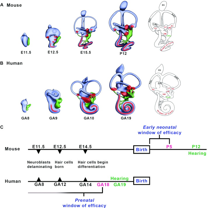
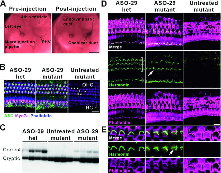
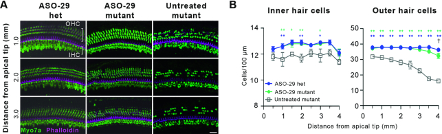
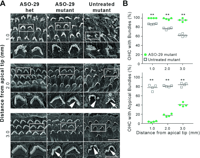
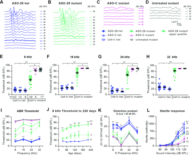
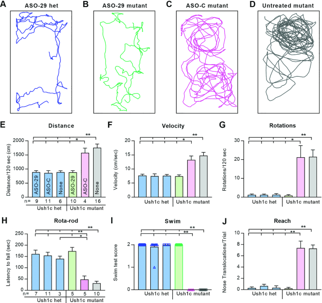
Disabling hearing loss impacts ∼466 million individuals worldwide with 34 million children affected. Gene and pharmacotherapeutic strategies to rescue auditory function in mouse models of human deafness are most effective when administered before hearing onset, after which therapeutic efficacy is significantly diminished or lost. We hypothesize that preemptive correction of a mutation in the fetal inner ear prior to maturation of the sensory epithelium will optimally restore sensory function. We previously demonstrated that transuterine microinjection of a splice-switching antisense oligonucleotide (ASO) into the amniotic cavity immediately surrounding the embryo on embryonic day 13-13.5 (E13-13.5) corrected pre-mRNA splicing in the juvenile Usher syndrome type 1c (Ush1c) mouse mutant. Here, we show that this strategy only marginally rescues hearing and partially rescues vestibular function. To improve therapeutic outcomes, we microinjected ASO directly into the E12.5 inner ear. A single intra-otic dose of ASO corrects harmonin RNA splicing, restores harmonin protein expression in sensory hair cell bundles, prevents hair cell loss, improves hearing sensitivity, and ameliorates vestibular dysfunction. Improvements in auditory and vestibular function were sustained well into adulthood. Our results demonstrate that an ASO pharmacotherapeutic administered to a developing organ system in utero preemptively corrects pre-mRNA splicing to abrogate the disease phenotype.


Figures

Figure 1.
Figure 1.
The onset of hearing in the postnatal mouse and the human fetus frames a window of therapeutic efficacy. (A, B) Mammalian inner ear morphogenesis. Lateral view of a right mouse (A) and human (B) inner ear from the otic vesicle stage to the elaboration of the membranous labyrinth. The onset of hearing defined by positive acoustic startle responses is first present at P12 in the mouse and at 19 weeks gestational age in the human (GA19; weeks since the first day of the last menstrual period) (29,30). Red, auditory and vestibular sensory patches; green, cochleovestibular ganglion. (C) Developmental timelines for mouse and human hearing onset. Noted are the approximate embryonic day (E) and weeks gestational age (GA) at which neurons and auditory sensory hair cells are born and differentiate. An early neonatal window of therapeutic efficacy in the mouse is present immediately after birth to P5 (3,6,7,9,10), one week prior to postnatal hearing onset (90). A corollary prenatal window of efficacy in humans is predicted to close by GA18, one week prior to fetal hearing onset (29,30). We hypothesize that fetal gene and pharmacotherapies will be required to achieve optimal restoration of sensory function in human inner ear diseases with congenital or early neonatal onset. Abbreviations: AC: anterior crista; ASC: anterior semicircular canal; CVG: cochleovestibular ganglion; E, embryonic day; ES, endolymphatic sac; LC, lateral crista; LSC, lateral semicircular canal; PC, posterior crista; PSC: posterior semicircular canal, SM, saccular macula; UM, utricular macula.
Figure 2.
Figure 2.
Transuterine microinjection of ASO-29 into the otic vesicle restores harmonin mRNA and protein expression. (A) ASO delivery by transuterine microinjection into the lumen of the left otic vesicle is validated by observation of the endolymphatic and cochlear ducts post-injection (compare to Figure 1A, E11.5). PHV: primary head vein. Scale bar: 1 mm. (B) ASO-29 localizes to inner hair cells (IHC, short bracket), outer hair cells (OHC, long bracket) as well as the Hensen's cell region (H, lateral to the third row of OHCs) and the pillar cell region (P, in between IHCs and the first row of OHCs) at P30. Asterisks indicate missing OHCs in the untreated mutant. Het: heterozygote. Scale bar: 20 μm. (C) Correctly spliced harmonin mRNA was detected in the P30 cochlea of ASO-29-treated heterozygous and mutant mice with a primer set that also detects the cryptically spliced form. (D) Harmonin protein expression was observed in stereociliary bundles of IHCs and OHCs of the cochlear apex in ASO-29-treated mutants at P30. Phalloidin labels filamentous actin. Arrow indicates a misshapen bundle in the ASO-29 treated mutant. Scale bar: 10 μm. (E) Higher magnification of OHCs showing harmonin expression in the bundles. Arrow indicates a misshapen bundle in the ASO-29 treated mutant. Scale bar: 5 μm. Fluorescent images are representative of n = 3 per treatment group in (B), (D) and (E). For splicing assessment, n = 4 per group in (C).
Figure 3.
Figure 3.
ASO-29 enhances inner and outer hair cell survival. (A) Representative whole mount Myosin7a (Myo7a) confocal microscopic projections showing enhanced hair cell survival at 1.0, 2.0 and 3.0 mm from the tip of the cochlear apex (8–24 kHz inclusive) in ASO-29-treated mutants at P30. Distance from the apical tip was translated to frequencies using the cochlear place frequency map shown in Supplementary Figure S2A. Scale bar: 20 μm. (B) Quantification of IHCs and OHCs ± SEM 0.5 to 4 mm from the tip of the cochlear apex (8–32 kHz inclusive). Untreated mutants were compared to the two ASO-29 treatment groups by two-way repeated measures ANOVA with Tukey's Multiple Comparison Test: *P < 0.05, **P < 0.01, n = 5 per group. The number of outer hair cells in ASO-29 mutants and ASO-29 heterozygotes was significantly different only at 4 mm by two-way repeated measures ANOVA with Tukey's Multiple Comparison Test: P < 0.05, n = 5 per group.
Figure 4.
Figure 4.
ASO-29 treatment improves outer hair cell stereociliary bundle morphology. (A) OHC stereociliary bundle morphology is improved in ASO-29-treated mutant inner ears 1.0, 2.0 and 3.0 mm from the tip of the cochlear apex (8–24 kHz inclusive) at P30. White boxes indicate the bundles shown in the subjacent high magnification panels. Arrows indicate misshapen bundles. Arrowheads indicate discontinuous bundles. Scale bars: 5 μm and 10 μm for the high and low magnification panels, respectively. (B) The percentage of OHCs with stereociliary bundles and with atypical bundles in ASO-29 treated mutant cochlea and untreated mutant cochleae at P30. Bundle presence and morphology were scored in 120 μm arcs from scanning electron micrographs taken at 1, 2 and 3 mm (8–24 kHz inclusive) from the tip of the cochlear apex. An atypical bundle displayed waviness or discontinuity as shown in panel (A). A total of 595 and 629 OHCs were scored in 4 ASO-29 treated mutants and four untreated mutants, respectively (**P < 0.01; two-tailed unpaired t-test).
Figure 5.
Figure 5.
ASO-29 rescues behaviorally-relevant auditory function that is sustained longitudinally. (A–D), ABR waveforms at 8 kHz from a representative of each of the four treatment groups. ABR thresholds are indicated by the thickened traces in (A) and (B); no thresholds were detected in the ASO-C treated mutant or the untreated mutant in (C) and (D). (E–H) Individual ABR thresholds at the four frequencies tested for every animal in the six treatment groups at P30. (I) ABR thresholds ± SEM for the cohorts in (E–H). The dotted line shows the average ABR thresholds ± SEM for seven ASO-29-treated animals in the upper (top-performing) quartile. (J) ABR thresholds ± SEM for 12 ASO-29-treated mutants followed through 220 days. Average thresholds for the upper quartile (n = 3) are significantly different from treated heterozygotes only at 140 days. (K) Distortion product otoacoustic emission (DPOAE) responses to an f2 stimulus of 60 dB SPL across frequencies ± SEM. The dotted line shows the average of 7 ASO-29-treated animals in the upper quartile (same subjects as in 5I, dotted line). n values in (E) apply to panels (F–I) and (K). (L) Acoustic startle reflex responses ± SEM. Statistical tests: panels (E–I) and (K): Kruskal–Wallis H Test with Dunn's Multiple Comparison Test comparing all groups to untreated Ush1c mutant; panel (J): repeated measures two-way ANOVA with Dunnett's Multiple Comparison Test comparing ASO-29-treated mutants to ASO-29-treated heterozygotes. (L), same as (J), but comparing all groups to untreated Ush1c mutant. *P < 0.05 and **P < 0.01. Level of significance for the comparisons involving each of the three heterozygous control groups is P < 0.01 and represented by a single asterisk pair in (I) and (K) (**) to reduce the visual complexity of the graph.
Figure 6.
Figure 6.
ASO-29 rescues behaviorally-relevant vestibular function that is sustained longitudinally. (A–D) Thirty second activity traces from a representative of each of the four treatment groups. (E–J) Graphs show average values ± SEM for: (E) distance traveled; (F) average velocity; (G) number of 360 degree rotations; (H) latency to fall; (I) swim score (individuals were plotted so that the zero scores of ASO-C and untreated mutants are readily detected on the X-axis); (J) average number of nose translocations past the shoulder line per trial (i.e., reaching caudally). One-way ANOVA with Tukey's Multiple Comparison Test; **P < 0.01 with comparisons indicated by the hatched bars above each panel. n values in (E) apply to (F) and (G); n values in (H) apply to (I) and (J). The 180 day vestibular function test data are presented in Supplementary Figure S9.

Similar articles

Cited by

References

    1. Olusanya B.O. Neonatal hearing screening and intervention in resource-limited settings: an overview. Arch. Dis. Child. 2012; 97:654–659. - PubMed
    1. Shearer A.E., Hildebrand M.S., Smith R.J.H.. Adam MP, Ardinger HH, Pagon RA, Wallace SE, Bean LJH, Mefford HC, Stephens K, Amemiya A, Ledbetter N.. 1993; Seattle (WA)GeneReviews®.
    1. Akil O., Seal R.P., Burke K., Wang C., Alemi A., During M., Edwards R.H., Lustig L.R.. Restoration of hearing in the VGLUT3 knockout mouse using virally mediated gene therapy. Neuron. 2012; 75:283–293. - PMC - PubMed
    1. Askew C., Rochat C., Pan B., Asai Y., Ahmed H., Child E., Schneider B.L., Aebischer P., Holt J.R.. Tmc gene therapy restores auditory function in deaf mice. Sci. Transl. Med. 2015; 7:295ra108. - PMC - PubMed
    1. Gyorgy B., Sage C., Indzhykulian A.A., Scheffer D.I., Brisson A.R., Tan S., Wu X., Volak A., Mu D., Tamvakologos P.I. et al. .. Rescue of hearing by gene delivery to Inner-Ear hair cells using Exosome-Associated AAV. Mol. Ther. 2017; 25:379–391. - PMC - PubMed

Publication types

MeSH terms