

Nanoparticle-Based Platform for Activatable Fluorescence Imaging and Photothermal Ablation of Endometriosis
- PMID: 32250034
- PMCID: PMC7210057
- DOI: 10.1002/smll.201906936
Nanoparticle-Based Platform for Activatable Fluorescence Imaging and Photothermal Ablation of Endometriosis
Abstract
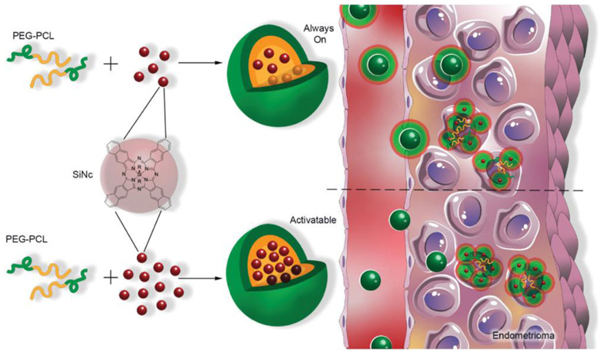
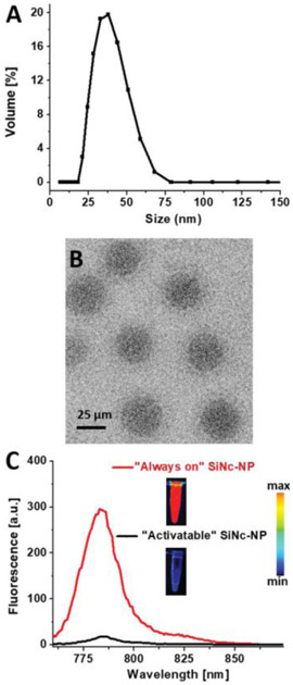
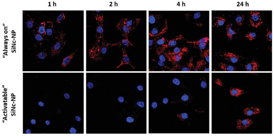
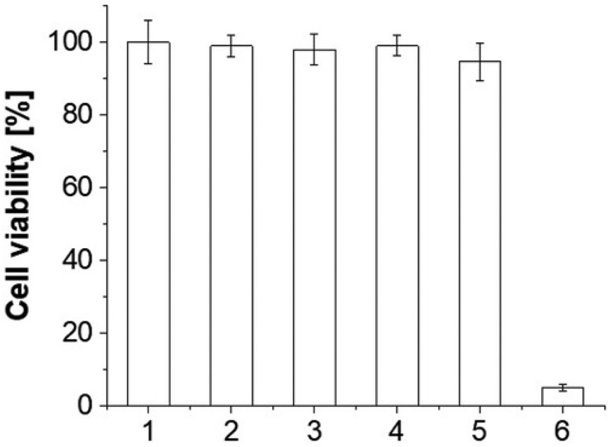
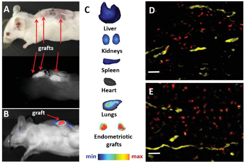
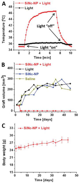
Endometriosis is a painful disorder where endometrium-like tissue forms lesions outside of the uterine cavity. Intraoperative identification and removal of these lesions are difficult. This study presents a nanoplatform that concurrently delineates and ablates endometriosis tissues using real-time near-infrared (NIR) fluorescence and photothermal therapy (PTT). The nanoplatform consists of a dye, silicon naphthalocyanine (SiNc), capable of both NIR fluorescence imaging and PTT, and a polymeric nanoparticle as a SiNc carrier to endometriosis tissue following systemic administration. To achieve high contrast during fluorescence imaging of endometriotic lesions, nanoparticles are constructed to be non-fluorescent prior to internalization by endometriosis cells. In vitro studies confirm that these nanoparticles activate the fluorescence signal following internalization in macaque endometrial stromal cells and ablate them by increasing cellular temperature to 53 ° C upon interaction with NIR light. To demonstrate in vivo efficiency of the nanoparticles, biopsies of endometrium and endometriosis from rhesus macaques are transplanted into immunodeficient mice. Imaging with the intraoperative Fluobeam 800 system reveals that 24 h following intravenous injection, nanoparticles efficiently accumulate in, and demarcate, endometriotic grafts with fluorescence. Finally, the nanoparticles increase the temperature of endometriotic grafts up to 47 °C upon exposure to NIR light, completely eradicating them after a single treatment.
Keywords: endometriosis; fluorescence imaging; nanoparticles; photothermal therapy.
© 2020 WILEY-VCH Verlag GmbH & Co. KGaA, Weinheim.
Conflict of interest statement
Conflict of Interest
The authors declare no conflict of interest.
Figures
Similar articles
-
A Tumor-Activatable Theranostic Nanomedicine Platform for NIR Fluorescence-Guided Surgery and Combinatorial Phototherapy.Theranostics. 2018 Jan 1;8(3):767-784. doi: 10.7150/thno.21209. eCollection 2018. Theranostics. 2018. PMID: 29344305 Free PMC article.
-
Dendrimer-encapsulated naphthalocyanine as a single agent-based theranostic nanoplatform for near-infrared fluorescence imaging and combinatorial anticancer phototherapy.Nanoscale. 2015 Mar 7;7(9):3888-902. doi: 10.1039/c4nr06050d. Nanoscale. 2015. PMID: 25422147
-
Targeted nanoparticles for imaging and therapy of endometriosis†.Biol Reprod. 2024 Jun 12;110(6):1191-1200. doi: 10.1093/biolre/ioae073. Biol Reprod. 2024. PMID: 38738758 Free PMC article. Review.
-
Assessing fluorescence detection and effective photothermal therapy of near-infrared polymer nanoparticles using alginate tissue phantoms.Lasers Surg Med. 2018 Dec;50(10):1040-1049. doi: 10.1002/lsm.22955. Epub 2018 Jun 28. Lasers Surg Med. 2018. PMID: 29953621 Free PMC article.
-
Emerging Trends in Nanotechnology for Endometriosis: Diagnosis to Therapy.Nanomaterials (Basel). 2024 Jun 5;14(11):976. doi: 10.3390/nano14110976. Nanomaterials (Basel). 2024. PMID: 38869601 Free PMC article. Review.
Cited by
-
Discovery and Validation of a Compound to Target Ewing's Sarcoma.Pharmaceutics. 2021 Sep 24;13(10):1553. doi: 10.3390/pharmaceutics13101553. Pharmaceutics. 2021. PMID: 34683845 Free PMC article.
-
Nanoparticle-Based Techniques for Bladder Cancer Imaging: A Review.Int J Mol Sci. 2023 Feb 14;24(4):3812. doi: 10.3390/ijms24043812. Int J Mol Sci. 2023. PMID: 36835222 Free PMC article. Review.
-
Targeted Nanoparticles with High Heating Efficiency for the Treatment of Endometriosis with Systemically Delivered Magnetic Hyperthermia.Small. 2022 Jun;18(24):e2107808. doi: 10.1002/smll.202107808. Epub 2022 Apr 17. Small. 2022. PMID: 35434932 Free PMC article.
-
Imaging of Endometriotic Lesions Using cRGD-MN Probe in a Mouse Model of Endometriosis.Nanomaterials (Basel). 2024 Feb 5;14(3):319. doi: 10.3390/nano14030319. Nanomaterials (Basel). 2024. PMID: 38334590 Free PMC article.
-
The Role of Nanomedicine in Benign Gynecologic Disorders.Molecules. 2024 May 1;29(9):2095. doi: 10.3390/molecules29092095. Molecules. 2024. PMID: 38731586 Free PMC article. Review.
References
Publication types
MeSH terms
Grants and funding
LinkOut - more resources
Full Text Sources
Medical
Miscellaneous
