Multicenter Study
. 2020 Mar 23;10(1):5199.
doi: 10.1038/s41598-020-62049-0.

Usefulness of diffusion tensor imaging findings as biomarkers for amyotrophic lateral sclerosis

Affiliations
Multicenter Study

Usefulness of diffusion tensor imaging findings as biomarkers for amyotrophic lateral sclerosis

Seol-Hee Baek et al. Sci Rep. .

Abstract

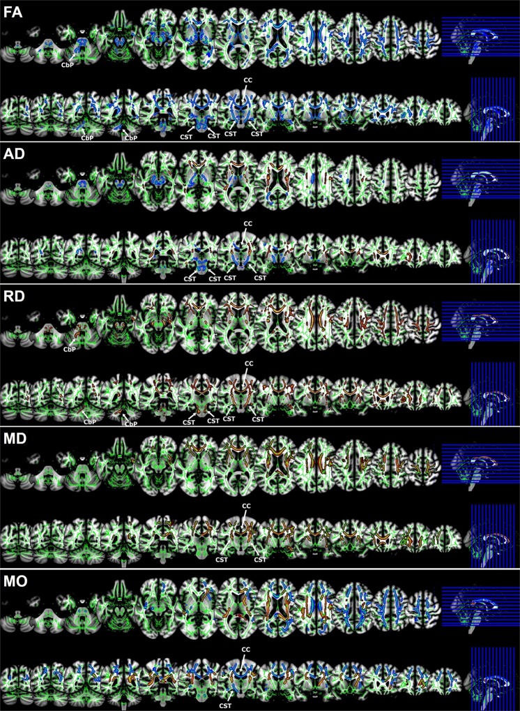
Amyotrophic lateral sclerosis (ALS) is a neurodegenerative disease. However, no reliable biomarkers have been identified to represent the clinical status. This study aimed to investigate whether diffusion tensor imaging (DTI) findings are useful imaging biomarkers to indicate the clinical status of ALS patients. Ninety-six probable or definite ALS cases and 47 age- and sex-matched, normal controls were enrolled. Demographic and clinical data were collected at the time of DTI. DTI data were acquired using a 3-Tesla magnetic resonance imaging scanner and analysed by voxel-wise statistical analyses for fractional anisotropy, axial diffusivity, radial diffusivity, mean diffusivity, and mode of anisotropy. Compared with the healthy control group, the ALS group had significant differences in DTI scalars in the diffuse tracts of the brain, which was predominant in the corticospinal tract at the brainstem and cerebellar peduncle area. Furthermore, the DTI values correlated with the ALS functional rating scale-revised (ALSFRS-R) scores and the delta ALSFRS-R score representing the rate of disease progression. The subgroup analysis revealed a more severe and widespread brain degeneration was observed in rapidly progressive ALS. Therefore, our results suggest that DTI findings are useful as imaging biomarkers for evaluating the clinical severity and rate of disease progression in ALS.


Conflict of interest statement

The authors declare no competing interests.

Figures

Figure 1
Figure 1
Flow chart of identification of patients with amyotrophic lateral sclerosis. We reviewed 109 patients with clinically probable or definite amyotrophic lateral sclerosis; and excluded 13 patients who manifested the first symptoms before 40 years of age. Subgroup analyses were performed according to the symptom onset regions, cognitive profiles, and the rate of disease progression. Only ALS patients without genetic mutations were included in subgroup analyses.
Figure 2
Figure 2
Tract-based spatial statistics: areas of significant differences in each diffusion tensor imaging parameter in amyotrophic lateral sclerosis patients compared with normal controls. Comparisons of diffusion tensor imaging between amyotrophic lateral sclerosis (ALS) and normal controls: fractional anisotropy (FA) maps showed the decreased FA value in the corpus callosum (CC), corona radiata (CR), cerebral peduncle (CP), corticospinal tract (CST), and cerebellar peduncle (CbP). On axial diffusivity (AD) maps, ALS had significantly decreased AD values in the bilateral CST at both the internal capsule and brainstem level, whereas there were significantly increased AD values in the CC and bilateral CR. Radial diffusivity (RD) and mean diffusivity (MD) values of ALS were increased in the CC and bilateral CR. In the mode of anisotropy (MO) image, ALS had significantly decreased MO values in the CC and left CST, whereas there were significantly increased MO values in the bilateral internal capsule.

References

    1. Rowland LP, Shneider NA. Amyotrophic lateral sclerosis. N. Engl. J. Med. 2001;344:1688–1700. doi: 10.1056/nejm200105313442207. - DOI - PubMed
    1. Zufiria M, et al. ALS: A bucket of genes, environment, metabolism and unknown ingredients. Prog. Neurobiol. 2016;142:104–129. doi: 10.1016/j.pneurobio.2016.05.004. - DOI - PubMed
    1. Ng AS, Rademakers R, Miller BL. Frontotemporal dementia: a bridge between dementia and neuromuscular disease. Ann. N. Y. Acad. Sci. 2015;1338:71–93. doi: 10.1111/nyas.12638. - DOI - PMC - PubMed
    1. Tae WS, Ham BJ, Pyun SB, Kang SH, Kim BJ. Current clinical applications of diffusion-tensor imaging in neurological disorders. J. Clin. Neurol. 2018;14:129–140. doi: 10.3988/jcn.2018.14.2.129. - DOI - PMC - PubMed
    1. Roberts RE, Anderson EJ, Husain M. White matter microstructure and cognitive function. The Neuroscientist. 2013;19:8–15. doi: 10.1177/1073858411421218. - DOI - PMC - PubMed

Publication types