Review
. 2021 Jan;26(1):296-308.
doi: 10.1038/s41380-020-0721-9. Epub 2020 Apr 6.

Biomarkers for Alzheimer's disease-preparing for a new era of disease-modifying therapies

Affiliations
Review

Biomarkers for Alzheimer's disease-preparing for a new era of disease-modifying therapies

Henrik Zetterberg et al. Mol Psychiatry. 2021 Jan.

Abstract

Clinical trial results presented in 2019 suggest that antibody-based removal of cerebral amyloid β (Aβ) plaques may possibly clear tau tangles and modestly slow cognitive decline in symptomatic Alzheimer's disease (AD). Although regulatory approval of this approach is still pending, preparing the healthcare system for the advent of disease-modifying therapies against AD is imperative. In particular, it will be necessary to identify the most suitable biomarkers to facilitate appropriate treatment of AD. Here, we give an update on recent developments in fluid and imaging biomarkers for AD-related pathologies and discuss potential approaches that could be adopted to screen for and clarify the underlying pathology in people seeking medical advice because of cognitive symptoms. We succinctly review recent data regarding biomarkers for Aβ and tau pathology, neurodegeneration, synaptic dysfunction, and inflammation, highlight the need for further research into common copathologies, and suggest how different biomarkers could be used (most likely in combination) to facilitate the development and clinical implementation of novel drug candidates against AD.


Conflict of interest statement

Conflicts of interest

HZ has served at scientific advisory boards for Denali, Roche Diagnostics, Wave, Samumed and CogRx, has given lectures in symposia sponsored by Fujirebio, Alzecure and Biogen, and is a co-founder of Brain Biomarker Solutions in Gothenburg AB, a GU Ventures-based platform company at the University of Gothenburg. BBB has received precursors and imaging agents from Avid Radiopharmaceuticals.

Figures
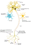
Figure 1.
Figure 1.
Biomarkers for Alzheimer’s disease. The figure illustrates neural cells and Alzheimer’s disease (AD) pathology, with AD-related biomarkers indicated in text boxes. Cerebrospinal fluid (CSF) and plasma Aβ42/Aβ40 ratio, as well as amyloid positron emission tomography (PET) are direct markers of Aβ pathology. In response to Aβ pathology, neurons phosphorylate and secrete tau at increased rate, resulting in increased total and phosphorylated tau (T-tau and P-tau, respectively) concentrations in CSF and in increased P-tau concentration in plasma. CSF and plasma tau may thus be considered neuronal response markers to Aβ. The most direct biomarker for tangle pathology is tau PET. CSF and plasma P-tau concentrations also increase in tau PET-positive individuals but the increase happens well before tau pathology is detectable on PET, at least using current tracers. Axons are rich in tau and neurofilament light (NfL) that leak into the CSF and blood during neuroaxonal degeneration. The best-established imaging biomarker for neurodegeneration is volumetric magnetic resonance imaging (MRI) of the brain. Leading synaptic biomarkers are neurogranin (Ng) in CSF, as well as SV2A- and fluorodeoxyglucose (FDG) PET. The best-established astrocytic biomarker is CSF YKL-40 and there are also promising data on CSF and plasma glial fibrillary acidic protein (GFAP) as an astrocytic activation/degeneration marker. The best-established biomarkers for microglia are CSF soluble triggering receptor expressed on myeloid cells 2 (sTREM2) and translocator protein (TSPO) PET.
Figure 2.
Figure 2.
Representative neuroimaging scans for assessing primary and secondary Alzheimer-related pathologies. (A) Warm/red color in the standardized uptake value ratio (SUVR) image denotes regions of amyloid deposition, as imaged with [11C] Pittsburgh Compound-B (PiB) positron emission tomography (PET) in an individual with Alzheimer’s disease (AD) dementia. (B) Warm/red color in the SUVR image denotes areas of [18F]AV1451-binding, indicating neurofibrillary tangle pathology in an individual with AD dementia. (C) Cooler colors (green/blue) are indicative of hypometabolism as shown on [18F] fluorodeoxyglucose (FDG) PET in an individual with mild cognitive impairment due to AD. (D) The compound [C11]UCB-J binds to synaptic vesicle glycoprotein 2A (SV2A). Red color in the SUVR image denotes regions of greater synaptic density in a healthy cognitively unimpaired (CU) late middle-aged adult.
Figure 3.
Figure 3.
A model of the temporal pattern of biomarker abnormalities for AD-related pathophysiological processes. The first biomarkers to change are cerebrospinal fluid (CSF) and plasma Aβ42/Aβ40 ratio. This is shortly followed by CSF and plasma tau increases, as a neuronal response to the amyloid changes. Shortly thereafter amyloid PET turns positive. Then microglia react and secrete soluble triggering receptor expressed on myeloid cells 2 (sTREM2), which reaches its maximum in the MCI stage of the disease, whereafter it declines in the dementia phase (there is not enough data on translocator protein PET, as a biomarker for microglial activation, to put it into the model). CSF neurogranin (Ng) is an early marker of synaptic dysfunction and increases in close association with amyloid PET positivity. When tau PET turns positive, a range of neurodegeneration and synaptic dysfunction biomarkers (CSF and serum/plama neurofilament light [NfL], hippocampal volume, and SV2A- and fluorodeoxyglucose [FDG] PET) change more or less in parallel. Most studies suggest that astrocytic biomarkers (YKL-40 and glial fibrillary acidic protein [GFAP]) change relatively late in the disease process.

References

    1. DeTure MA, Dickson DW. The neuropathological diagnosis of Alzheimer's disease. Mol Neurodegener. 2019;14:32. - PMC - PubMed
    1. Palmqvist S, Insel PS, Stomrud E, Janelidze S, Zetterberg H, Brix B, et al. Cerebrospinal fluid and plasma biomarker trajectories with increasing amyloid deposition in Alzheimer's disease. EMBO Mol Med. 2019;11:e11170. - PMC - PubMed
    1. Jack CR Jr., Holtzman DM. Biomarker modeling of Alzheimer's disease. Neuron. 2013;80:1347–58. - PMC - PubMed
    1. Hardy JA, Higgins GA. Alzheimer's disease: the amyloid cascade hypothesis. Science. 1992;256:184–5. - PubMed
    1. De Strooper B, Karran E. The Cellular Phase of Alzheimer's Disease. Cell. 2016;164:603–15. - PubMed

Publication types

MeSH terms