Review
. 2020 Jul;21(7):398-414.
doi: 10.1038/s41580-020-0232-1. Epub 2020 Apr 6.

The clearance of dead cells by efferocytosis

Affiliations
Review

The clearance of dead cells by efferocytosis

Emilio Boada-Romero et al. Nat Rev Mol Cell Biol. 2020 Jul.

Abstract

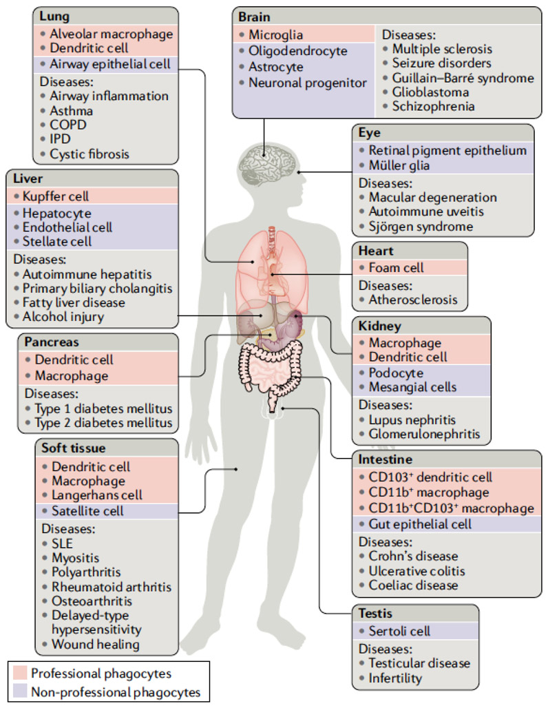
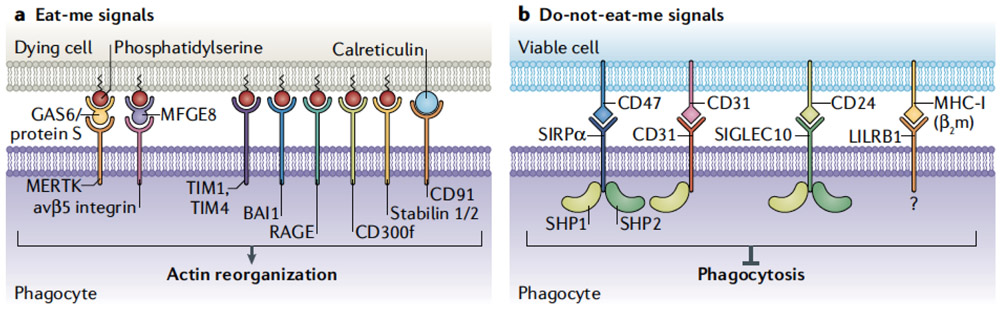
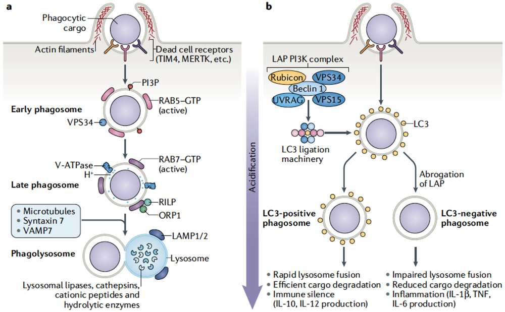
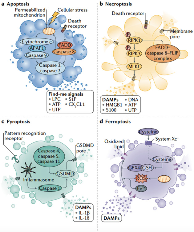
Multiple modes of cell death have been identified, each with a unique function and each induced in a setting-dependent manner. As billions of cells die during mammalian embryogenesis and daily in adult organisms, clearing dead cells and associated cellular debris is important in physiology. In this Review, we present an overview of the phagocytosis of dead and dying cells, a process known as efferocytosis. Efferocytosis is performed by macrophages and to a lesser extent by other 'professional' phagocytes (such as monocytes and dendritic cells) and 'non-professional' phagocytes, such as epithelial cells. Recent discoveries have shed light on this process and how it functions to maintain tissue homeostasis, tissue repair and organismal health. Here, we outline the mechanisms of efferocytosis, from the recognition of dying cells through to phagocytic engulfment and homeostatic resolution, and highlight the pathophysiological consequences that can arise when this process is abrogated.


Figures

Figure 1.
Figure 1.. Efferocytosis is critical for tissue homeostasis.
Efferocytosis can be carried out by professional phagocytes (red boxes), such as macrophages and dendritic cells, or to a lesser extent by non-professional phagocytes (blue boxes) such as epithelial cells. Disruption of normal efferocytosis can contribute to the development of a wide range of pathologies (light grey boxes) across a variety of tissues. (dark grey boxes). COPD, chronic obstructive pulmonary disease; IPD, idiopathic pulmonary disease; SLE, systemic lupus erythematosus.
Figure 2.
Figure 2.. Cell surface signals regulating efferocytosis.
Cells display proteins and lipids to communicate with phagocytes. These protein signals can be “eat-me” signals or “don’t eat me” signals, and interact with phagocytes via receptors on the phagocyte cell membrane. A) Dying cells expose a variety of signals on their surface that serve as “eat-me” signals for surveying phagocytes. A primary signal exposed on dying cells is the externalization of phosphatidylserine (PS). PS can interact with a variety of receptors on the surface of the phagocyte including TIM1, TIM4, BAI1, RAGE, the CD300 family, and the Stabilin proteins. Additionally, signaling can occur through receptors including MerTK, via adaptors including Gas6 and protein S, or through avβ5 integrin via MFGE-8. Calreticulin expressed on the surface of the dying cell can act as an eat-me signal through binding to CD91 expressed on the phagocyte outer membrane. B) Healthy cells decorate their plasma membranes with “don’t-eat-me” signals that protect them from phagocytic targeting. Don’t-eat-me signals include CD47 which is recognized by SIRPα on the phagocyte surface, and CD31, which homodimerizes with CD31 on phagocytes. Similarly, CD24 on viable cells engages Siglec-10 on phagocytes to inhibit engulfment. Signaling in each case occurs through SHP1/2 and suppression of pathways required for phagocytosis, including actin remodeling. Class I MHC (MHC-I) can signal through LILRB1 to mitigate phagocytic activation, although the signaling cascade is not fully delineated. In combination, these signals regulate the initiation of efferocytosis.
Figure 3.
Figure 3.. Phagocyte processing of a dying cell.
Upon engulfment by a phagocyte, a dying cell progresses through a canonical program leading to the degradation of the dead cell by the lysosome. Additional modifying pathways can be engaged that alter the degradative process and subsequent downstream immune signaling pathways. A) Efferocytosis of dying cells is a multistep process, involving initiation of phagocytosis, maturation of the phagosome, degradation of phagolysosome contents, and the resultant downstream effects on physiological outcomes. B) The process of LC3-associated phagocytosis (LAP) is characterized by recruitment of LC3 to the phagosome membrane. Following engagement of phagocytosis, a PI3-kinase complex containing Rubicon, UVRAG, Beclin1, VPS34, and VPS15, is assembled at the phagophore. Assembly of this PI3-kinase complex leads to the recruitment and activation of the LC3 ligation machinery. Ligation of LC3 to the phagosome lipid membrane promotes rapid maturation and lysosomal fusion. This expeditious process leads to efficient clearance of the dead cell and immune silence. If LAP is abrogated, there is a delay in phagosome maturation and lysosomal fusion. The impaired clearance of the dying cell leads to inflammatory immune activation through processes that are, to date, not fully understood.
Box 1 Figure
Box 1 Figure

References

    1. Galluzzi L et al. Molecular mechanisms of cell death: recommendations of the Nomenclature Committee on Cell Death 2018. Cell Death Differ 25, 486–541, doi:10.1038/s41418-017-0012-4 (2018).

      A useful set of definitions of diverse modes of cell death based on mechanisms rather than morphology.

    1. Bergmann A & Steller H Apoptosis, stem cells, and tissue regeneration. Sci Signal 3, re8, doi:10.1126/scisignal.3145re8 (2010). - DOI - PMC - PubMed
    1. Suzanne M & Steller H Shaping organisms with apoptosis. Cell Death Differ 20, 669–675, doi:10.1038/cdd.2013.11 (2013). - DOI - PMC - PubMed
    1. Fuchs Y & Steller H Programmed cell death in animal development and disease. Cell 147, 742–758, doi:10.1016/j.cell.2011.10.033 (2011). - DOI - PMC - PubMed
    1. Blander JM Death in the intestinal epithelium-basic biology and implications for inflammatory bowel disease. FEBS J 283, 2720–2730, doi: 10.1111/febs.13771 (2016). - DOI - PMC - PubMed

Publication types