. 2020 May;17(5):481-494.
doi: 10.1038/s41592-020-0796-x. Epub 2020 Apr 6.

Genetic tool development in marine protists: emerging model organisms for experimental cell biology

Drahomíra Faktorová #  1 R Ellen R Nisbet #  2   3 José A Fernández Robledo #  4 Elena Casacuberta #  5 Lisa Sudek #  6 Andrew E Allen  7   8 Manuel Ares Jr  9 Cristina Aresté  5 Cecilia Balestreri  10 Adrian C Barbrook  2 Patrick Beardslee  11 Sara Bender  12 David S Booth  13 François-Yves Bouget  14 Chris Bowler  15 Susana A Breglia  16 Colin Brownlee  10 Gertraud Burger  17 Heriberto Cerutti  11 Rachele Cesaroni  18 Miguel A Chiurillo  19 Thomas Clemente  11 Duncan B Coles  4 Jackie L Collier  20 Elizabeth C Cooney  21 Kathryn Coyne  22 Roberto Docampo  19 Christopher L Dupont  8 Virginia Edgcomb  23 Elin Einarsson  2 Pía A Elustondo  16   24 Fernan Federici  25 Veronica Freire-Beneitez  26   27 Nastasia J Freyria  4 Kodai Fukuda  28 Paulo A García  29 Peter R Girguis  30 Fatma Gomaa  30 Sebastian G Gornik  31 Jian Guo  6   9 Vladimír Hampl  32 Yutaka Hanawa  33 Esteban R Haro-Contreras  16 Elisabeth Hehenberger  21 Andrea Highfield  10 Yoshihisa Hirakawa  33 Amanda Hopes  34 Christopher J Howe  2 Ian Hu  2 Jorge Ibañez  25 Nicholas A T Irwin  21 Yuu Ishii  35 Natalia Ewa Janowicz  32 Adam C Jones  12 Ambar Kachale  36 Konomi Fujimura-Kamada  37 Binnypreet Kaur  36 Jonathan Z Kaye  12 Eleanna Kazana  26   27 Patrick J Keeling  21 Nicole King  13 Lawrence A Klobutcher  38 Noelia Lander  19 Imen Lassadi  2 Zhuhong Li  19 Senjie Lin  38 Jean-Claude Lozano  14 Fulei Luan  11 Shinichiro Maruyama  35 Tamara Matute  25 Cristina Miceli  39 Jun Minagawa  37   40 Mark Moosburner  7   8 Sebastián R Najle  5   41 Deepak Nanjappa  22 Isabel C Nimmo  2 Luke Noble  42   43 Anna M G Novák Vanclová  32 Mariusz Nowacki  18 Isaac Nuñez  25 Arnab Pain  44   45 Angela Piersanti  39 Sandra Pucciarelli  39 Jan Pyrih  26   32 Joshua S Rest  46 Mariana Rius  20 Deborah Robertson  47 Albane Ruaud  25   48 Iñaki Ruiz-Trillo  5   49   50 Monika A Sigg  13 Pamela A Silver  51   52 Claudio H Slamovits  16 G Jason Smith  53 Brittany N Sprecher  38 Rowena Stern  10 Estienne C Swart  18   48 Anastasios D Tsaousis  26   27 Lev Tsypin  54   55 Aaron Turkewitz  54 Jernej Turnšek  7   8   51   52 Matus Valach  17 Valérie Vergé  14 Peter von Dassow  25   56 Tobias von der Haar  26 Ross F Waller  2 Lu Wang  57 Xiaoxue Wen  11 Glen Wheeler  10 April Woods  53 Huan Zhang  38 Thomas Mock  58 Alexandra Z Worden  59   60 Julius Lukeš  61
Affiliations

Genetic tool development in marine protists: emerging model organisms for experimental cell biology

Drahomíra Faktorová et al. Nat Methods. 2020 May.

Erratum in

  • Publisher Correction: Genetic tool development in marine protists: emerging model organisms for experimental cell biology.
    Faktorová D, Nisbet RER, Fernández Robledo JA, Casacuberta E, Sudek L, Allen AE, Ares M Jr, Aresté C, Balestreri C, Barbrook AC, Beardslee P, Bender S, Booth DS, Bouget FY, Bowler C, Breglia SA, Brownlee C, Burger G, Cerutti H, Cesaroni R, Chiurillo MA, Clemente T, Coles DB, Collier JL, Cooney EC, Coyne K, Docampo R, Dupont CL, Edgcomb V, Einarsson E, Elustondo PA, Federici F, Freire-Beneitez V, Freyria NJ, Fukuda K, García PA, Girguis PR, Gomaa F, Gornik SG, Guo J, Hampl V, Hanawa Y, Haro-Contreras ER, Hehenberger E, Highfield A, Hirakawa Y, Hopes A, Howe CJ, Hu I, Ibañez J, Irwin NAT, Ishii Y, Janowicz NE, Jones AC, Kachale A, Fujimura-Kamada K, Kaur B, Kaye JZ, Kazana E, Keeling PJ, King N, Klobutcher LA, Lander N, Lassadi I, Li Z, Lin S, Lozano JC, Luan F, Maruyama S, Matute T, Miceli C, Minagawa J, Moosburner M, Najle SR, Nanjappa D, Nimmo IC, Noble L, Novák Vanclová AMG, Nowacki M, Nuñez I, Pain A, Piersanti A, Pucciarelli S, Pyrih J, Rest JS, Rius M, Robertson D, Ruaud A, Ruiz-Trillo I, Sigg MA, Silver PA, Slamovits CH, Jason Smith G, Sprecher BN, Stern R, Swart EC, Tsaousis AD, Tsypin L, Turkewitz A, Turnšek J, Valach M, Vergé V, von Dassow P, von der Haar T, Waller RF, Wa… See abstract for full author list ➔ Faktorová D, et al. Nat Methods. 2020 May;17(5):551. doi: 10.1038/s41592-020-0828-6. Nat Methods. 2020. PMID: 32296171 Free PMC article.

Abstract

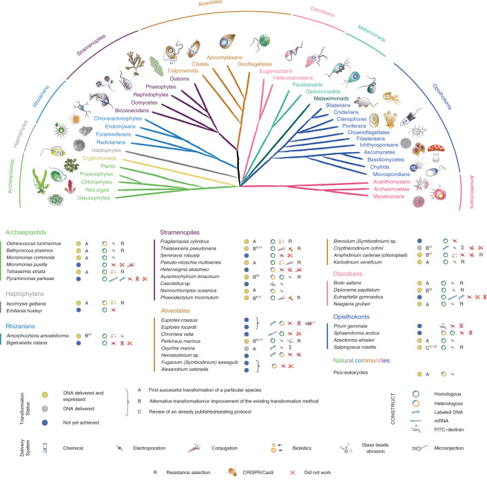
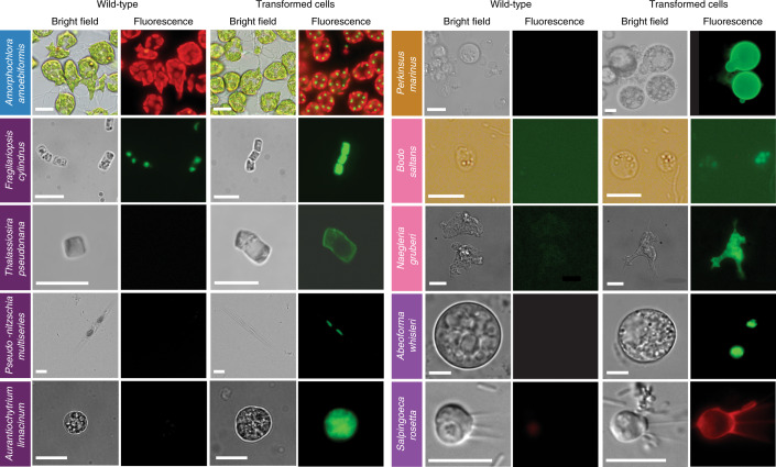
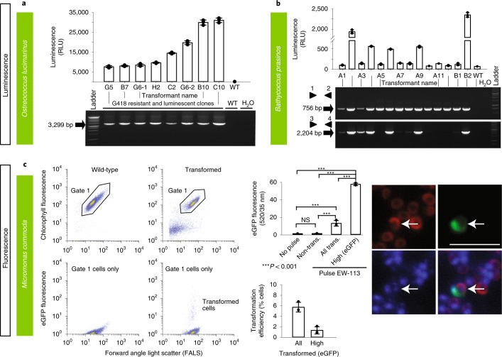
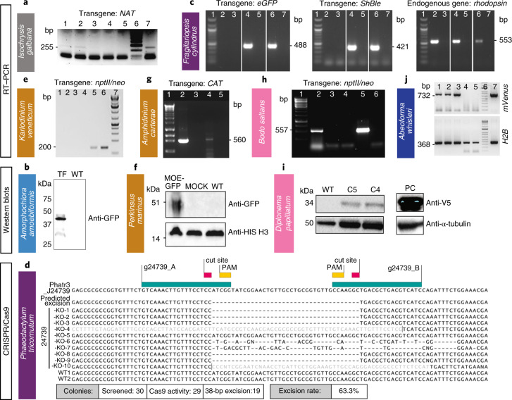
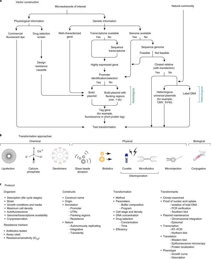
Diverse microbial ecosystems underpin life in the sea. Among these microbes are many unicellular eukaryotes that span the diversity of the eukaryotic tree of life. However, genetic tractability has been limited to a few species, which do not represent eukaryotic diversity or environmentally relevant taxa. Here, we report on the development of genetic tools in a range of protists primarily from marine environments. We present evidence for foreign DNA delivery and expression in 13 species never before transformed and for advancement of tools for eight other species, as well as potential reasons for why transformation of yet another 17 species tested was not achieved. Our resource in genetic manipulation will provide insights into the ancestral eukaryotic lifeforms, general eukaryote cell biology, protein diversification and the evolution of cellular pathways.


Conflict of interest statement

The authors declare no competing interests.

Figures

Fig. 1
Fig. 1. Phylogenetic relationships and transformation status of marine protists.
A schematic view of the eukaryotic tree of life with effigies of main representatives. Color-coordinated species we have attempted to genetically modify are listed below. Current transformability status is schematized in circles indicating: DNA delivered and shown to be expressed (yellow, for details see text and Table 1); DNA delivered, but no expression seen (gray) and no successful transformation achieved despite efforts (blue). The details of transformation of species that belong to ‘DNA delivered’ and ‘Not achieved yet’ categories are described in Supplementary Table 5. mRNA, messenger RNA; FITC–dextran, fluorescein isothiocyanate (FITC)-conjugated dextran.
Fig. 2
Fig. 2. Epifluorescence micrographs of transformed marine protists.
Representative images of transformants and wild-type cell lines of ten selected protist species. Colored boxes behind species names refer to phylogenetic supergroup assignments given in Fig. 1. Representative data of at least two independent experiments are shown. The fluorescent images show the expression of individual fluorescent marker genes introduced via transformation for all organisms shown, except in the case of A. amoebiformis. For this, red depicts the natural autofluorescence of photosynthetic pigments in the cell, while the additional green spheres in the transformant fluorescence panel shows introduced GFP fluorescence (see Supplementary Fig. 15c for a trace of these different regions in the cell). Scale bars are as follows: 10 µm for A. amoebiformis, T. pseudonana, A. limacinum, B. saltans, N. gruberi, A. whisleri and S. rosetta; 15 µm for P. marinus; 20 µm for F. cylindrus and 100 µm for P. multiseries.
Fig. 3
Fig. 3. Various methods were used to demonstrate successful transformation in different archaeplastid species: luminescence and fluorescence.
ac, Luminescence (a,b) and fluorescence (by FACS and epifluorescence microscopy) (c) were used to verify expression of introduced constructs in three archaeplastids: O. lucimarinus (a), B. prasinos (b) and M. commoda (c). For the latter, red in the image depicts the natural autofluorescence of photosynthetic pigments in the cell, while green shows introduced eGFP fluorescence and blue shows the DAPI-stained nucleus; the overlay shows colocalization of eGFP and nucleus signals. See Supplementary Fig. 15d for a trace of these different regions in the cell. NS, not significant; trans., transformed. Representative data of at least two independent experiments are shown. For a detailed figure description see Supplementary Notes 2. Source data
Fig. 4
Fig. 4. Various methods were used to demonstrate successful transformation in different species: RT–PCR, western blot and sequencing.
aj, Western blot, RT–PCR or sequencing (in case of Cas9-induced excision by CRISPR) were used to verify expression of introduced constructs in one haptophyte: I. galbana (a), one rhizarian—A. amoebiformis (b), two stramenopiles—F. cylindrus (c) and P. tricornutum (d), three alveolates—K. veneficum (e), P. marinus (f) and A. carterae (g), two discobans—B. saltans (h) and D. papillatum (i) and one opisthokont—A. whisleri (j). Note that nptII/neo is used synonymously with amino 3′-glycosyl phosphotransferase gene (aph(3′)) conferring resistance to kanamycin and neomycin. Representative data of at least two independent experiments are shown. For a detailed figure description see Supplementary Notes 2. Source data
Fig. 5
Fig. 5. ‘Transformation roadmap’ for the creation of genetically tractable protists.
a, Vector design and construction for microeukaryotes of interest and a natural community. b, Transformation approaches. Different symbols represent methods (for example chemical, physical or biological) for introducing DNA/RNA/protein into a living cell. c, Protocol. Key methodological steps for successful transformation are listed in an abbreviated form (for particular examples, see Table 1 and text).

Comment in

  • A genetic toolbox for marine protists.
    Brown MW, Tice AK. Brown MW, et al. Nat Methods. 2020 May;17(5):469-470. doi: 10.1038/s41592-020-0794-z. Nat Methods. 2020. PMID: 32251395 No abstract available.

References

    1. Worden AZ, et al. Rethinking the marine carbon cycle: factoring in the multifarious lifestyles of microbes. Science. 2015;347:1257594. - PubMed
    1. de Vargas C, et al. Eukaryotic plankton diversity in the sunlit global ocean. Science. 2015;348:1261605. - PubMed
    1. Collier JL, Rest JS. Swimming, gliding, and rolling toward the mainstream: cell biology of marine protists. Mol. Biol. Cell. 2019;30:1245–1248. - PMC - PubMed
    1. Curtis BA, et al. Algal genomes reveal evolutionary mosaicism and the fate of nucleomorphs. Nature. 2012;492:59–65. - PubMed
    1. Armbrust EV, et al. The genome of the diatom Thalassiosira pseudonana: ecology, evolution, and metabolism. Science. 2004;306:79–86. - PubMed

Publication types