. 2020 Apr 6;18(4):e3000704.
doi: 10.1371/journal.pbio.3000704. eCollection 2020 Apr.

Single-cell mapping reveals new markers and functions of lymphatic endothelial cells in lymph nodes

Affiliations

Single-cell mapping reveals new markers and functions of lymphatic endothelial cells in lymph nodes

Noriki Fujimoto et al. PLoS Biol. .

Abstract

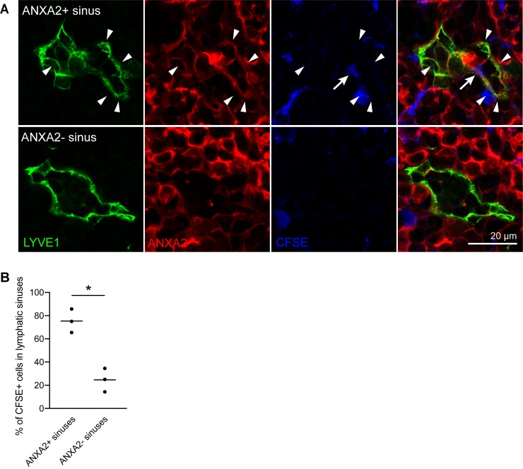
Lymph nodes (LNs) are highly organized secondary lymphoid organs that mediate adaptive immune responses to antigens delivered via afferent lymphatic vessels. Lymphatic endothelial cells (LECs) line intranodal lymphatic sinuses and organize lymph and antigen distribution. LECs also directly regulate T cells, mediating peripheral tolerance to self-antigens, and play a major role in many diseases, including cancer metastasis. However, little is known about the phenotypic and functional heterogeneity of LN LECs. Using single-cell RNA sequencing, we comprehensively defined the transcriptome of LECs in murine skin-draining LNs and identified new markers and functions of distinct LEC subpopulations. We found that LECs residing in the subcapsular sinus (SCS) have an unanticipated function in scavenging of modified low-density lipoprotein (LDL) and also identified a specific cortical LEC subtype implicated in rapid lymphocyte egress from LNs. Our data provide new, to our knowledge, insights into the diversity of LECs in murine LNs and a rich resource for future studies into the regulation of immune responses by LN LECs.


Conflict of interest statement

The authors have declared that no competing interests exist.

Figures

Fig 1
Fig 1. Single-cell transcriptomic analysis of LN LECs.
(A) Example FACS plot of LN stromal cells (pregated as CD45- living singlets) from inguinal LNs of C57Bl/6 wild-type mice. CD31+Pdpn+ LECs were isolated by single-cell sorting. (B) Unsupervised clustering of 893 LN LECs resulted in 4 distinct clusters. Each point represents an individual cell. (C, D) Expression levels of selected genes plotted using the original log-transformed counts. Gray dots indicate cells without any measurable expression; red dots coded by color intensity denote the detected expression magnitude. (C) Pecam1 (CD31) and Pdpn used as markers for FACS sorting, as well as the LEC marker genes Prox1 and Flt4 (Vegfr3), were robustly expressed in most cells. (D) While Lyve1 and Itga2b were expressed in all clusters except for cluster 2, the fLEC marker Madcam1 and the cLEC marker Ackr4 were specifically expressed in cluster 1 and cluster 2, respectively. ACKR4, atypical chemokine receptor 4; CD, cluster of differentiation; cLEC, ceiling LEC; FACS, fluorescence-activated cell sorting; fLEC, floor-lining LEC; Flt4, Fms related receptor tyrosine kinase 4; ITGA2B, integrin subunit alpha 2b; LEC, lymphatic endothelial cell; LN, lymph node; LYVE1, lymphatic vessel endothelial hyaluronan receptor 1; MADCAM1, mucosal vascular addressin cell adhesion molecule 1; Pdpn, podoplanin; Pecam1, platelet and endothelial cell adhesion molecule 1; Prox1, prospero homeobox 1; UMAP, Uniform Manifold Approximation and Projection; VEGF, vascular endothelial growth factor.
Fig 2
Fig 2. Molecular characterization of LECs in the SCS floor.
(A) Immunofluorescence staining of a whole inguinal LN cross-section for IgD (green), CD3 (red), and LYVE1 (white). The dotted squares indicate 3 regions that were used for further analysis: the SCS (SCS associated with B cell follicles), the IF-SCS (SCS and large IF sinus tracts entering the node), and the MS within the node. (B) Immunofluorescence costaining for LYVE1 (green) and ITGA2B (red) in the SCS (upper panels) and the IF-SCS region (lower panels) showed expression of ITGA2B and LYVE1 by LECs in both the SCS floor and the IF-SCS region. (C–E) Gene expression patterns (left panels; denoted as high expression level in red and low in blue, using the corrected expression values) and protein/transcript location of new fLEC (cluster 1) markers. CD44 (C, detected by immunofluorescence staining), Glycam1 (D, detected by RNA FISH), and Coch (E, detected by RNA FISH) were specifically expressed in fLECs in the SCS region (upper right panels), but not in the IF-SCS region (lower right panels). White arrowheads indicate RNA FISH signals; LYVE1 stained in green. CD, cluster of differentiation; cLEC, ceiling LEC; Coch, cochlin; FISH, fluorescence in situ hybridization; fLEC, floor-lining LEC; Glycam1, glycosylation-dependent cell adhesion molecule 1; IF, interfollicular; IgD, immunoglobulin D; ITGA2B, integrin subunit alpha 2b; LEC, lymphatic endothelial cell; LN, lymph node; LYVE1, lymphatic vessel endothelial hyaluronan receptor 1; MS, medullary sinus; SCS, subcapsular sinus; UMAP, Uniform Manifold Approximation and Projection.
Fig 3
Fig 3. Molecular characterization of LECs in the SCS ceiling with immunofluorescence staining.
(A–C) Expression of new cLEC/cluster 2 marker genes ANXA2 (A), FABP4 (B), and CD36 (C) by RNA sequencing (left panels) and immunofluorescence staining (right panels) in Ackr4-GFP reporter mice. GFP (white) and immunofluorescence costaining for LYVE1 (green) served as markers for cLECs and fLECs, respectively. ACKR4, atypical chemokine receptor 4; ANXA2, annexin A2; CD, cluster of differentiation; cLEC, ceiling LEC; FABP4, fatty acid binding protein 4; fLEC, floor-lining LEC; GFP, green fluorescent protein; LEC, lymphatic endothelial cell; LYVE1, lymphatic vessel endothelial hyaluronan receptor 1; SCS, subcapsular sinus; UMAP, Uniform Manifold Approximation and Projection.
Fig 4
Fig 4. Molecular characterization of LECs in the SCS ceiling with RNA FISH.
(A-B) Expression of new cLEC/cluster 2 marker genes Ackr3 (A) and Btnl9 (B) by RNA sequencing (left panels) and RNA FISH (right panels). As GFP fluorescence is lost during tissue processing for RNA FISH, immunofluorescence staining for ANXA2 (red) and LYVE1 (green) served as markers for cLECs and fLECs, respectively. Arrows point to cLECs expressing Ackr3 and Btnl9 transcripts (white). ACKR3, atypical chemokine receptor 3; ANXA2, annexin A2; Btnl9, butyrophilin like 9; cLEC, ceiling LEC; FISH, fluorescence in situ hybridization; fLEC, floor-lining LEC; LEC, lymphatic endothelial cell; LYVE1, lymphatic vessel endothelial hyaluronan receptor 1; SCS, subcapsular sinus; UMAP, Uniform Manifold Approximation and Projection.
Fig 5
Fig 5. Differential LDL uptake by cLECs.
In vivo LDL tracing after intradermal injection of Dil-labeled acetylated or oxidized LDL near the base of the tail of Ackr4-GFP reporter mice. Draining inguinal LNs were collected 1 h later. (A, B) Representative images (A) and quantification (B) of acetylated LDL (red) accumulation in ACKR4+ and LYVE1+ LECs. Each line represents one mouse (n = 6). (C–D) Representative images (C) and quantification (D) of oxidized LDL (red) accumulation in ACKR4+ and LYVE1+ LECs. Empty arrowheads indicate cLECs, and filled arrowheads indicated fLECs. For quantification, signal intensities were normalized to the level of ACKR4- LYVE1+ LECs (n = 5). **p < 0.01; *p < 0.05 (paired t test). For raw quantitative data, please refer to S1 Data. ACKR4, atypical chemokine receptor 4; A.U., arbitrary unit; cLEC, ceiling LEC; Dil, 1,1′-dioctadecyl-3,3,3′,3′-tetramethylindocarbocyanine perchlorate; fLEC, floor-lining LEC; GFP, green fluorescent protein; LDL, low-density lipoprotein; LEC, lymphatic endothelial cell; LN, lymph node; LYVE1, lymphatic vessel endothelial hyaluronan receptor 1.
Fig 6
Fig 6. Molecular characterization of MS LECs.
(A–C) Expression of MS LEC/cluster 3 marker genes IL33 (A), MRC1 (B), and MARCO (C) by RNA sequencing (left panels) and immunofluorescence staining (right panels, red) in combination with LYVE1 (green). IL33 (A) was expressed by fLECs and MS LECs and showed nuclear LEC staining in the SCS, IF-SCS, and MS regions, whereas MRC1 (B) and MARCO (C) were excluded from fLECs and congruously showed no staining in the SCS region. fLEC, floor-lining LEC; IF, interfollicular; IL33, interleukin 33; LEC, lymphatic endothelial cell; LYVE1, lymphatic vessel endothelial hyaluronan receptor 1; MARCO, macrophage receptor with collagenous structure; MRC1, mannose receptor C-type 1; MS, medullary sinus; SCS, subcapsular sinus; UMAP, Uniform Manifold Approximation and Projection.
Fig 7
Fig 7. A unique subset of cortical and MSs.
(A) Representative images of immunofluorescence staining for LYVE1 (green) and ANXA2 (red), in combination with RNA FISH to detect Glycam1 (white). ANXA2+ lymphatic sinuses are present in the cortex, close to the medulla (indicated by white arrowheads), and are often in close proximity to Glycam1+ HEVs (indicated by asterisks). (B–D) Expression of the cluster 4 LEC marker genes Ptx3 (B), Kcnj8 (C), and Itih5 (D) by RNA sequencing (left panels) and RNA FISH (right panels). Immunofluorescence staining for ANXA2 (red) and LYVE1 (green) was used to highlight cluster 4 sinuses. White arrowheads point to LECs expressing Ptx3, Kcnj8, and Itih5 transcripts (white). ANXA2, annexin A2; FISH, fluorescence in situ hybridization; Glycam1, glycosylation-dependent cell adhesion molecule 1; HEV, high endothelial venule; Itih5, inter-alpha-trypsin inhibitor heavy chain 5; Kcnj8, potassium inwardly rectifying channel subfamily J member 8; LYVE1, lymphatic vessel endothelial hyaluronan receptor 1; MS, medullary sinus; Ptx3, pentraxin 3; UMAP, Uniform Manifold Approximation and Projection.
Fig 8
Fig 8. Lymphocytes egress from LNs via cluster 4 sinuses.
CFSE-labeled splenocytes were infused via the tail vein, and inguinal LNs were collected 30 min later. (A) Representative images of an ANXA2+ sinus (top) and an ANXA2- sinus (bottom), stained for LYVE1 (green) and ANXA2 (red). Arrowheads indicate ANXA2+ LECs. CFSE-labeled splenocytes (blue) eventually entered lymphatic sinuses (arrow). (B) Quantification of CFSE+ splenocytes that entered ANXA2+ cluster 4 sinuses or other (ANXA2-) lymphatic sinuses 30 min after infusion, expressed as percentage of splenocytes that entered any kind of lymphatic sinus. CFSE-labeled cells were more frequently observed in ANXA2+ cluster 4 sinuses than in ANXA2- medullary sinuses. Each symbol represents one mouse (n = 3). *p < 0.05 (paired t test). For raw quantitative data, please refer to S1 Data. ANXA2, annexin A2; CFSE, carboxy-fluorescein diacetate succinimidyl ester; LEC, lymphatic endothelial cell; LN, lymph node; LYVE1, lymphatic vessel endothelial hyaluronan receptor 1.
Fig 9
Fig 9. Overview of murine LN LEC subsets and comparison to human LN LECs.
(A–C) Gene-level comparison between mouse and human [23] fLECs (A), cLECs (B), and medullary LECs (C), based on up-regulated (top row) and down-regulated (bottom) genes in each cluster compared to all other LECs. Venn diagrams display the number of differentially expressed genes that are shared or different between the 2 data sets. (D) Expression of the selected markers (x-axis) signifying individual LN LEC subsets (y-axis). Dot size denotes the proportion of cells with detectable expression. Color intensity indicates the relative mean expression level of the corresponding gene, using the original log-transformed counts. For raw quantitative data, please refer to S1 Data. ACKR4, atypical chemokine receptor 4; ANXA2, annexin A2; Bgn, biglycan; CD, cluster of differentiation; cLEC, ceiling LEC; Coch, cochlin; FABP4, fatty acid binding protein 4; fLEC, floor-lining LEC; Flrt2, fibronectin leucine-rich transmembrane protein 2; Glycam1, glycosylation-dependent cell adhesion molecule 1; IL33, interleukin 33; ITGA2B, integrin subunit alpha 2b; Itih5, inter-alpha-trypsin inhibitor heavy chain 5; Kcnj8, potassium inwardly rectifying channel subfamily J member 8; LEC, lymphatic endothelial cell; LN, lymph node; LYVE1, lymphatic vessel endothelial hyaluronan receptor 1; MARCO, macrophage receptor with collagenous structure; MRC1, mannose receptor C-type 1; Ptx3, pentraxin 3.

References

    1. Clement CC, Wang W, Dzieciatkowska M, Cortese M, Hansen KC, Becerra A, et al. Quantitative Profiling of the Lymph Node Clearance Capacity. Sci Rep. 2018;8(1):11253 10.1038/s41598-018-29614-0 - DOI - PMC - PubMed
    1. Rantakari P, Auvinen K, Jappinen N, Kapraali M, Valtonen J, Karikoski M, et al. The endothelial protein PLVAP in lymphatics controls the entry of lymphocytes and antigens into lymph nodes. Nat Immunol. 2015;16(4):386–96. 10.1038/ni.3101 . - DOI - PubMed
    1. Reynoso GV, Weisberg AS, Shannon JP, McManus DT, Shores L, Americo JL, et al. Lymph node conduits transport virions for rapid T cell activation. Nat Immunol. 2019;20(5):602–12. 10.1038/s41590-019-0342-0 - DOI - PMC - PubMed
    1. Cyster JG, Schwab SR. Sphingosine-1-phosphate and lymphocyte egress from lymphoid organs. Annu Rev Immunol. 2012;30:69–94. Epub 2011 Dec 5. 10.1146/annurev-immunol-020711-075011 . - DOI - PubMed
    1. Cohen JN, Guidi CJ, Tewalt EF, Qiao H, Rouhani SJ, Ruddell A, et al. Lymph node-resident lymphatic endothelial cells mediate peripheral tolerance via Aire-independent direct antigen presentation. J Exp Med. 2010;207(4):681–8. 10.1084/jem.20092465 - DOI - PMC - PubMed

Publication types

LinkOut - more resources