. 2020 Apr 21;59(15):1493-1507.
doi: 10.1021/acs.biochem.9b01035. Epub 2020 Apr 13.

Biochemical Characterization of Yeast Xrn1

Affiliations

Biochemical Characterization of Yeast Xrn1

Conner J Langeberg et al. Biochemistry. .

Abstract

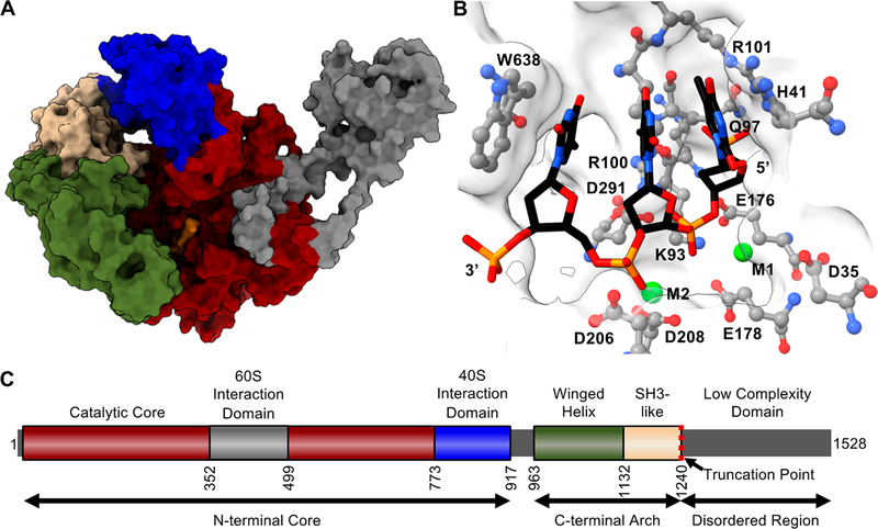
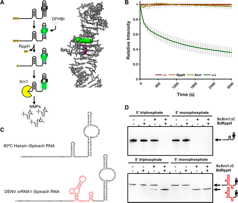
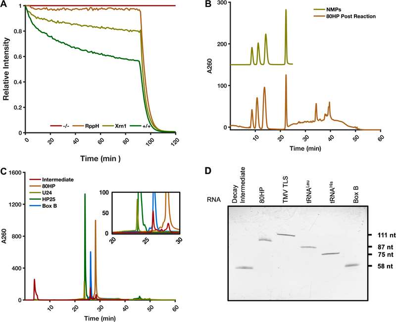
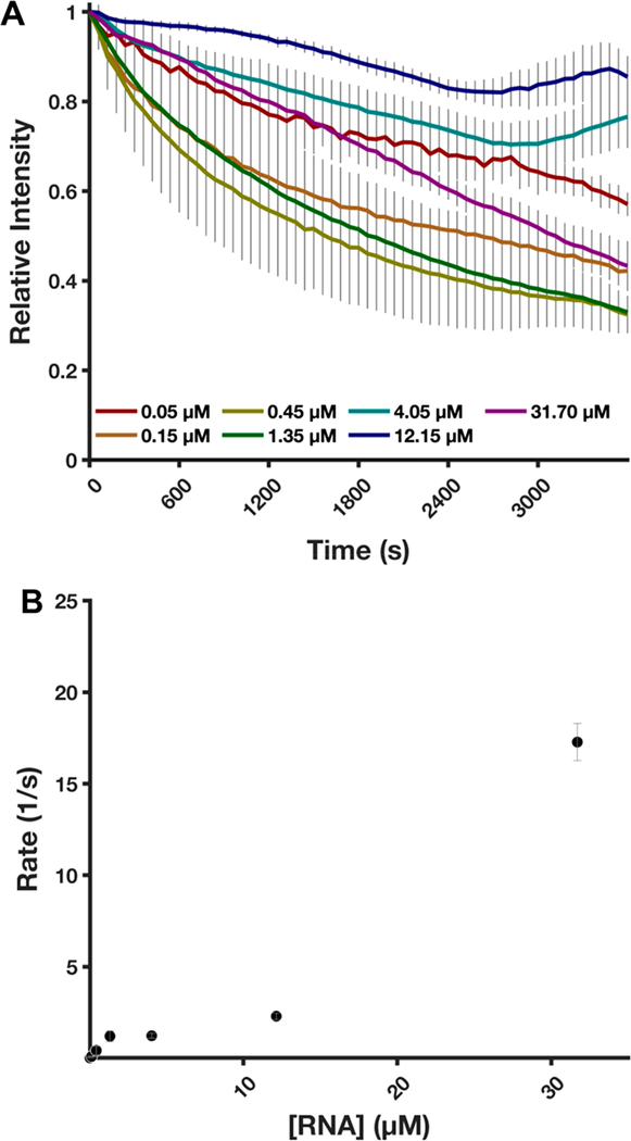
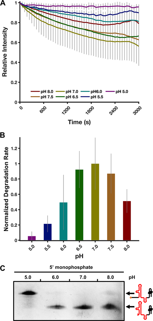
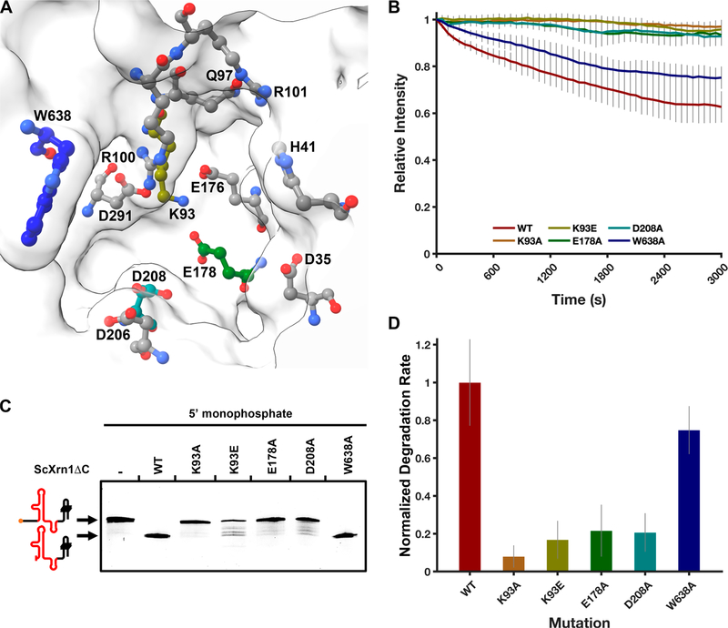
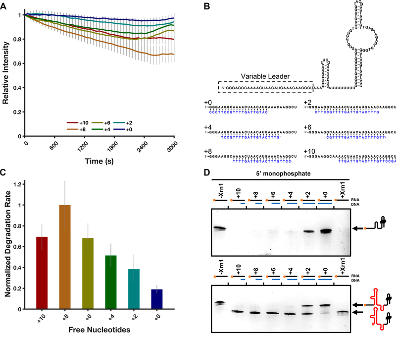
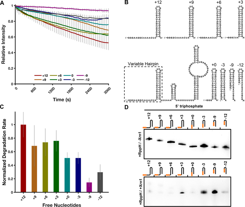
Messenger RNA degradation is an important component of overall gene expression. During the final step of eukaryotic mRNA degradation, exoribonuclease 1 (Xrn1) carries out 5' → 3' processive, hydrolytic degradation of RNA molecules using divalent metal ion catalysis. To initiate studies of the 5' → 3' RNA decay machinery in our lab, we expressed a C-terminally truncated version of Saccharomyces cerevisiae Xrn1 and explored its enzymology using a second-generation, time-resolved fluorescence RNA degradation assay. Using this system, we quantitatively explored Xrn1's preference for 5'-monophosphorylated RNA substrates, its pH dependence, and the importance of active site mutations in the molecule's conserved catalytic core. Furthermore, we explore Xrn1's preference for RNAs containing a 5' single-stranded region both in an intermolecular hairpin structure and in an RNA-DNA hybrid duplex system. These results both expand and solidify our understanding of Xrn1, a centrally important enzyme whose biochemical properties have implications in numerous RNA degradation and processing pathways.


Conflict of interest statement

The authors declare no competing financial interest.

Figures

Figure 1.
Figure 1.
Structural model of ScXrn1ΔC. (A) Structural model of ScXrn1ΔC generated using the I-TASSER iterative threading model. Regions are colored to correspond to domain architecture in panel C; a DNA trinucleotide substrate, dTdTdT, from the Drosophila structure (PDB entry 2Y35) is colored orange. (B) Active site of the I-TASSER-generated structure of ScXrn1ΔC with the TTT trinucleotide substrate from the Drosophila structure (PDB entry 2Y35) and metal ions from both Drosophila and Kluveromyces (PDB entry 3PIF) structures modeled in. (C) Domain architecture of ScXrn1. The N-terminal core (residues 1−773) of the enzyme includes the active site. The following region of residues 774−917 forms a domain recently shown to interact with the 40S subunit of the ribosome. Residues 963−1132 and 1133−1240 form motifs most closely resembling winged-helix and SH3 motifs, respectively. The truncation point of ScXrn1ΔC is indicated by a red dashed line at residue 1240.
Figure 2.
Figure 2.
Second-generation time-resolved, fluorescent RNA degradation assay. (A) Diagram of RNA processing in the TRFRD assays and an inset structure of the DFHBI-bound iSpinach aptamer (PDB entry 4TS2). DFHBI is colored green, and K+ ions are colored purple. (B) Normalized fluorescence trace of a TRFRD assay using the 80HP construct. The “−/−” trace shown in red is a no enzyme control. The “+/+” trace shown in green contains both RppH and ScXrn1ΔC. Conditions: 2 μM 80HP RNA, 2 μM BdRppH, and 1 μM ScXrn1ΔC in 1xEC3K+ buffer at pH 7.9 and 37 °C. Error bars represent one standard deviation for nine replicates. (C) Secondary structures of the two main RNA constructs used in this study, 80HP and DENVxrRNA1. The Xrn1-resistant structure is colored red. The layout of each construct is described in the text. (D) dPAGE of RNA constructs demonstrating the phosphorylation state dependency of ScXrn1ΔC. Conditions: 0.5 μM RNA, 0.5 μM BdRppH, and 0.25 μM ScXrn1ΔC in 1xEC3K+ buffer at pH 7.9 and 37 °C for 2 h.
Figure 3.
Figure 3.
Interrogation of ScXrn1ΔC products. (A) Typical TRFRD assay using ScXrn1ΔC with the addition of RNase A at 90 min. The “−/−” trace shown in red is a no enzyme control. The “+/+” trace shown in green contains both RppH and ScXrn1ΔC. Conditions: 2 μM 80HP RNA, 2 μM BdRppH, 0.05 μM ScXrn1ΔC, and 5 units of RNase A in 1xEC3K+ buffer at pH 7.9 and 37 °C. (B) HPLC trace of products from bulk reaction of 80HP RNA with ScXrn1ΔC (bottom trace). The top trace is the four purified 5′NMPs resolved using TBAP-modified C-18 chromatography. (C) HPLC analysis of the 80HP RNA with and without ScXrn1ΔC in the presence of DFHBI using PLRP-S chromatography overlaid with the trace of three oligonucleotide-length markers: U24 (24 nucleotides), HP25 (25 nucleotides), and Box B (58 nucleotides). The inset is a region of the trace where the DFHBI-dependent resistant RNA population appears during the reaction. We have not yet fully characterized the intermediate products, but their size roughly corresponds to the size of the iSpinach aptamer, indicating degradation up to the quadruplex DFHBI binding pocket. (D) dPAGE of the intermediate compared against the full-length 80HP construct and various RNA constructs as size markers. The intermediate appears to be approximately the same size as the 58-nucleotide Box B RNA.
Figure 4.
Figure 4.
ScXrn1ΔC kinetics. (A) Normalized trace of the TRFRD assay obtained during kinetic experiments varying the concentration of 5′-monophosphorylated RNA as indicated. The relative stoichiometries of ScXrn1ΔC molecules to RNA molecules are as follows: 1:1, 1:3, 1:9, 1:27, 1:81, 1:243, and 1:634. Conditions: 0.05 μM ScXrn1ΔC, 5′-monophosphorylated 80HP RNA concentrations as shown, 1xEC3K+ buffer, pH 7.9, 37 °C. (B) Quantification of rates of per phosphodiester bond hydrolysis. An enzyme-limited regime, or saturation, was not achieved using RNA concentrations of ≤31.7 μM. From the data, a lower bound for ScXrn1ΔC activity can be estimated to be 17.3 ± 0.6 s−1. Error reported as one standard deviation for nine replicates excluding 31.7 μM where n = 3.
Figure 5.
Figure 5.
ScXrn1ΔC exhibits pH-dependent activity. (A) Normalized trace of the TRFRD assay conducted using 80HP RNA and ScXrn1ΔC at different pHs. Conditions: 2 μM purified, 5′-monophosphorylated 80HP RNAs, and 0.05 μM ScXrn1ΔC in 1xEC3K+ buffer at the indicated pH and 37 °C. (B) Relative rates of ScXrn1ΔC-catalyzed degradation of 80HP RNA at the indicated pHs. Error reported as one standard deviation for nine replicates. (C) dPAGE of the DENVxrRNA1 RNA constructs showing the pH dependency of ScXrn1ΔC. Conditions: 0.5 μM RNA, 0.5 μM BdRppH, and 0.25 μM ScXrn1ΔC in 1xEC3K+ buffer at the indicated pH and 37 °C for 2 h.
Figure 6.
Figure 6.
pH titration of 5′-phosphate analogues, methyl phosphate and β-glycerol phosphate. (A) Quantum mechanical calculation of the surface potential of isobutyl phosphate showing the potential difference of the monoanionic (left) and dianionic (right) phosphate groups, calculated using Gaussian 09 at the B3LYP/6–31+G(d,p) level. (B) pH titration of a 10 mM methyl/dimethyl phosphate solution with 100 mM sodium hydroxide. pKa2 is indicated by a dashed line at pH 6.3. The inset scheme shows the relevant protonation/deprotonation reaction being monitored. (C) pH titration of a 10 mM β-glycerol phosphate solution with 100 mM phosphoric acid. pKa2 is indicated by a dashed line at pH 6.3. Again, the inset scheme depicts the relevant equilibrium.
Figure 7.
Figure 7.
ScXrn1ΔC activity is dependent on a set of conserved active site residues. (A) Model of the ScXrn1ΔC active site generated using the ITASSER iterative threading model. Previously mutagenized residues implicated in the mechanism of Xrn1 are shown: Asp35, His41, Lys93, Gln97, Arg100, Arg101, Glu176, Glu178, Asp206, Asp208, Asp291, and Trp638. Carbon atoms of the corresponding residues tested in this study are colored to correspond to the plots and bar graphs in this figure. (B) Normalized TRFRD assay traces of both WT and mutant ScXrn1ΔC constructs using the 80HP RNA. Conditions: 2 μM purified, 5′-monophosphorylated 80HP RNA, and 0.05 μM ScXrn1ΔC in 1xEC3K+ buffer at pH 7.9 and 37 °C. (C) dPAGE of both the wild type and mutants of ScXrn1ΔC using the DENVxrRNA1 construct. Conditions: 0.5 μM DENVxrRNA1 RNA and 0.25 μM ScXrn1ΔC in 1xEC3K+ buffer at pH 7.9 and 37 °C for 2 h. (D) Relative rates of RNA degradation carried out by the WT and mutant ScXrn1ΔC’s K93A, K93E, E178A, D208A, and W638A. Error reported as one standard deviation for nine replicates.
Figure 8.
Figure 8.
ScXrn1ΔC prefers RNAs with single-stranded ends. (A) Normalized TRFRD traces for degradation of DNA primer-annealed 80HP constructs by ScXrn1ΔC. Conditions: 2 μM purified, 5′-monophosphorylated 80HP RNA annealed to 2 μM indicated primer, and 0.05 μM ScXrn1ΔC in 1xEC3K+ buffer at pH 7.9 and 37 °C. (B) Secondary structure of the 80HP construct used in these experiments showing the indicated primers (blue) used to duplex the 5′ single-stranded region. (C) Relative rates of degradation for constructs with the indicated number of free 5′-nucleotides. Error reported as one standard deviation for nine replicates. (D) dPAGE of the 80HP and DENVxrRNA1 RNA constructs in panel B showing the single-stranded leader length dependency of ScXrn1ΔC. Conditions: 0.5 μM RNA, 0.5 μM BdRppH, and 0.25 μM ScXrn1ΔC in 1xEC3K+ buffer at pH 7.9 and 37 °C for 30 min.
Figure 9.
Figure 9.
ScXrn1ΔC prefers RNAs with exposed 5′-ends. (A) Normalized trace of the TRFRD assay using a buried hairpin RNA and ScXrn1ΔC. Conditions: 2 μM purified, 5′-triphosphorylated buried hairpin RNA as indicated, and 0.05 μM ScXrn1ΔC in 1xEC3K+ buffer at pH 7.9 and 37 °C. (B) Secondary structure of the buried hairpin constructs used in these experiments showing the indicated truncations made to the 5′ single-stranded region. (C) Relative rates of degradation for the indicated constructs. Error reported as one standard deviation for nine replicates. (D) Single-time point dPAGE analysis of the RNA constructs in panel B. Conditions: 0.5 μM RNA, 0.5 μM BdRppH, and 0.25 μM ScXrn1ΔC in 1xEC3K+ buffer at pH 7.9 and 37 °C for 30 min. Top, no ScXrn1ΔC added (controls). Bottom, reactions with both BdRppH and ScXrn1ΔC included.

References

    1. Parker R, and Song H (2004) The enzymes and control of eukaryotic mRNA turnover. Nat. Struct. Mol. Biol. 11, 121–127. - PubMed
    1. Garneau NL, Wilusz J, and Wilusz CJ (2007) The highways and byways of mRNA decay. Nat. Rev. Mol. Cell Biol. 8, 113–126. - PubMed
    1. Lebreton A, Tomecki R, Dziembowski A, and Séraphin B (2008) Endonucleolytic RNA cleavage by a eukaryotic exosome. Nature 456, 993–996. - PubMed
    1. Fabian MR, Sonenberg N, and Filipowicz W (2010) Regulation of mRNA Translation and Stability by microRNAs. Annu. Rev. Biochem. 79, 351–379. - PubMed
    1. Larimer FW, Hsu CL, Maupin MK, and Stevens A (1992) Characterization of the XRN1 gene encoding a 5′→3′ exoribonuclease: sequence data and analysis of disparate protein and mRNA levels of gene-disrupted yeast cells. Gene 120, 51–57. - PubMed

MeSH terms

Substances