Single-day Postnatal Alcohol Exposure Induces Apoptotic Cell Death and Causes long-term Neuron Loss in Rodent Thalamic Nucleus Reuniens
- PMID: 32251710
- PMCID: PMC7236664
- DOI: 10.1016/j.neuroscience.2020.03.046
Single-day Postnatal Alcohol Exposure Induces Apoptotic Cell Death and Causes long-term Neuron Loss in Rodent Thalamic Nucleus Reuniens
Abstract
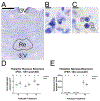
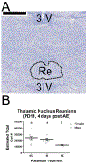
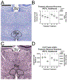
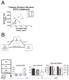
Fetal alcohol spectrum disorders (FASD) constitute a prevalent, yet preventable, developmental disorder worldwide. While a wealth of research demonstrates that altered function of hippocampus (HPC) and prefrontal cortex may underlie behavioral impairments in FASD, only one published paper to date has examined the impact of developmental alcohol exposure (AE) on the region responsible for coordinated prefrontal-hippocampal activity: thalamic nucleus reuniens (Re). In the current study, we used a rodent model of human third trimester AE to examine both the acute and lasting impact of a single-day AE on Re. We administered 5.25 g/kg of ethanol to male and female Long Evans rat pups on postnatal day (PD) 7. We used unbiased stereological estimation to evaluate cell death or cell loss at three time points: 12 h after alcohol administration; 4 days after alcohol administration (i.e., PD11); in adulthood (i.e.,PD 72). AE on PD7 increased apoptotic cell death in Re on PD7, and caused short-term cell loss on PD11. This relationship between short-term cell death versus cell number suggests that alcohol-related cell loss is driven by induction of apoptosis. In adulthood, alcohol-exposed animals displayed permanent cell loss (mediating volume loss in the Re), which included a reduction in neuron number (relative to procedural controls). Both procedural controls and alcohol exposed animals displayed a deficit in non-neuronal cell number relative to typically-developing controls, suggesting that Re cell populations may be vulnerable to early life stress as well as AE in an insult- and cell type-dependent manner.
Keywords: fetal alcohol spectrum disorders; histology; immunocytochemistry; neuroanatomy; stereology.
Copyright © 2020 IBRO. Published by Elsevier Ltd. All rights reserved.
Figures


Similar articles
-
Executive functioning-specific behavioral impairments in a rat model of human third trimester binge drinking implicate prefrontal-thalamo-hippocampal circuitry in Fetal Alcohol Spectrum Disorders.Behav Brain Res. 2021 May 7;405:113208. doi: 10.1016/j.bbr.2021.113208. Epub 2021 Feb 25. Behav Brain Res. 2021. PMID: 33640395 Free PMC article.
-
Rat Model of Late Gestational Alcohol Exposure Produces Similar Life-Long Changes in Thalamic Nucleus Reuniens Following Moderate- Versus High-Dose Insult.Alcohol Alcohol. 2022 Jul 9;57(4):413-420. doi: 10.1093/alcalc/agac008. Alcohol Alcohol. 2022. PMID: 35258554 Free PMC article.
-
Changes in Representation of Thalamic Projection Neurons within Prefrontal-Thalamic-Hippocampal Circuitry in a Rat Model of Third Trimester Binge Drinking.Brain Sci. 2021 Mar 4;11(3):323. doi: 10.3390/brainsci11030323. Brain Sci. 2021. PMID: 33806485 Free PMC article.
-
Nucleus reuniens of the midline thalamus of a rat is specifically damaged after early postnatal alcohol exposure.Neuroreport. 2019 Jul 3;30(10):748-752. doi: 10.1097/WNR.0000000000001270. Neuroreport. 2019. PMID: 31095109 Free PMC article.
-
Representation of prefrontal axonal efferents in the thalamic nucleus reuniens in a rodent model of fetal alcohol exposure during third trimester.Front Behav Neurosci. 2022 Sep 8;16:993601. doi: 10.3389/fnbeh.2022.993601. eCollection 2022. Front Behav Neurosci. 2022. PMID: 36160686 Free PMC article.
Cited by
-
Executive functioning-specific behavioral impairments in a rat model of human third trimester binge drinking implicate prefrontal-thalamo-hippocampal circuitry in Fetal Alcohol Spectrum Disorders.Behav Brain Res. 2021 May 7;405:113208. doi: 10.1016/j.bbr.2021.113208. Epub 2021 Feb 25. Behav Brain Res. 2021. PMID: 33640395 Free PMC article.
-
Midline Thalamic Damage Associated with Alcohol-Use Disorders: Disruption of Distinct Thalamocortical Pathways and Function.Neuropsychol Rev. 2021 Sep;31(3):447-471. doi: 10.1007/s11065-020-09450-8. Epub 2020 Aug 12. Neuropsychol Rev. 2021. PMID: 32789537 Free PMC article. Review.
-
Postnatal alcohol exposure and adolescent exercise have opposite effects on cerebellar microglia in rat.Int J Dev Neurosci. 2020 Oct;80(6):558-571. doi: 10.1002/jdn.10051. Epub 2020 Aug 4. Int J Dev Neurosci. 2020. PMID: 32681672 Free PMC article.
-
Upregulation of mesencephalic astrocyte-derived neurotrophic factor (MANF) expression offers protection against alcohol neurotoxicity.J Neurochem. 2023 Sep;166(6):943-959. doi: 10.1111/jnc.15921. Epub 2023 Jul 28. J Neurochem. 2023. PMID: 37507360 Free PMC article.
-
Longitudinal Evaluation Using Preclinical 7T-Magnetic Resonance Imaging/Spectroscopy on Prenatally Dose-Dependent Alcohol-Exposed Rats.Metabolites. 2023 Apr 6;13(4):527. doi: 10.3390/metabo13040527. Metabolites. 2023. PMID: 37110185 Free PMC article.
References
-
- Altman J and Bayer SA (1979). “Development of the diencephalon in the rat. IV. Quantitative study of the time of origin of neurons and the internuclear chronological gradients in the thalamus.” J Comp Neurol 188(3): 455–471. - PubMed
-
- Bates D, Machler M, Bolker B and Walker S (2015). “Fitting Linear Mixed-Effects Models Using lme4.” Journal of Statistical Software 67(1): 1–48.
-
- Berman RF and Hannigan JH (2000). “Effects of prenatal alcohol exposure on the hippocampus: spatial behavior, electrophysiology, and neuroanatomy.” Hippocampus 10(1): 94–110. - PubMed
Publication types
MeSH terms
Grants and funding
LinkOut - more resources
Full Text Sources
Medical

