Review
. 2020 Aug:72:109626.
doi: 10.1016/j.cellsig.2020.109626. Epub 2020 Apr 3.

Structure and function of polycystin channels in primary cilia

Affiliations
Review

Structure and function of polycystin channels in primary cilia

Chau My Ta et al. Cell Signal. 2020 Aug.

Abstract

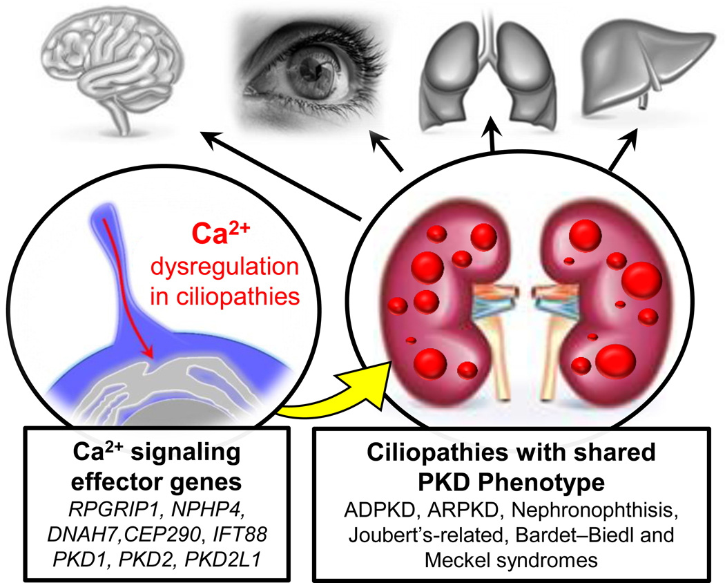
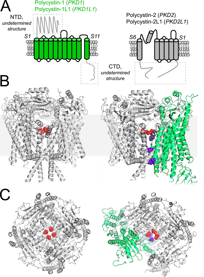
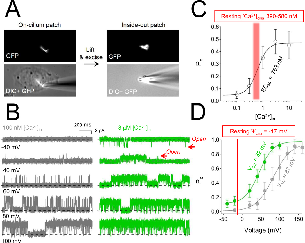
Variants in genes which encode for polycystin-1 and polycystin-2 cause most forms of autosomal dominant polycystic disease (ADPKD). Despite our strong understanding of the genetic determinants of ADPKD, we do not understand the structural features which govern the function of polycystins at the molecular level, nor do we understand the impact of most disease-causing variants on the conformational state of these proteins. These questions have remained elusive because polycystins localize to several organelle membranes, including the primary cilia. Primary cilia are microtubule based organelles which function as cellular antennae. Polycystin-2 and related polycystin-2 L1 are members of the transient receptor potential (TRP) ion channel family, and form distinct ion channels in the primary cilia of disparate cell types which can be directly measured. Polycystin-1 has both ion channel and adhesion G-protein coupled receptor (GPCR) features-but its role in forming a channel complex or as a channel subunit chaperone is undetermined. Nonetheless, recent polycystin structural determination by cryo-EM has provided a molecular template to understand their biophysical regulation and the impact of disease-causing variants. We will review these advances and discuss hypotheses regarding the regulation of polycystin channel opening by their structural domains within the context of the primary cilia.


Figures

Figure 1.
Figure 1.. Ciliary calcium dysregulation as a mechanism connecting the shared PKD phenotype found in several congenital ciliopathies.
Left, examples of calcium effector genes which encode for cilia-localized proteins. Right, the associated ciliopathies which primarily impact specific organs (in grey) but share PKD as common co-morbidity.
Figure 2.
Figure 2.. Topology and structure of hetero and homomeric polycystins.
A) Left, the topology of the eleven transmembrane proteins, polycystin-1 and polycystin-1L1 (green). Right, the topology of the six transmembrane polycystin-2 (PKD2) and polycystin-2L1 (PKD2L1) TRP channels. The boxed areas of the proteins have not been structurally solved. B) Transmembrane and C) extracellular structural views of the homomeric polycystin-2 channel (PDB: 5K47, Grieben et al 2017) and heteromeric polycystin-1 + polycystin-2 complex (PDB: 6A70, Su et al 2019). The polycystin-1 positively charged pore residues are colored purple, whereas the negatively charged polycystin-2 selectivity filter residues are colored red.
Figure 3.
Figure 3.. Calcium-dependent potentiation of polycystin-2 channels in the primary cilia.
A) Images of the inside out cilia patch configuration. B) Here, the inner membrane of the cilia faces the external solution, so that calcium concentrations can be adjusted while measuring singe channel activity while holding the membrane potential at different voltages. C) Polycystin-2 open probability (Po) and intraciliary calcium concentration relationship. The resting ciliary calcium concentration (red range) has been measured at 390 nM and 580 nM for RPE and IMCD cells, respectively,. D) The voltage dependence of polycystin-2 channel opening captured at low and high intra-ciliary calcium. The resting membrane potential of the cilia is shown in red, indicating that most of the polycytin-2 channels are closed when intraciliary calcium is low.

Similar articles

Cited by

References

    1. Harris PC & Rossetti S Molecular diagnostics for autosomal dominant polycystic kidney disease. Nat Rev Nephrol 6, 197–206, doi:10.1038/nrneph.2010.18 (2010). - DOI - PMC - PubMed
    1. Chebib FT & Torres VE Autosomal Dominant Polycystic Kidney Disease: Core Curriculum 2016. Am J Kidney Dis 67, 792–810, doi:10.1053/j.ajkd.2015.07.037 (2016). - DOI - PMC - PubMed
    1. Hughes J et al. The polycystic kidney disease 1 (PKD1) gene encodes a novel protein with multiple cell recognition domains. Nat Genet 10, 151–160, doi:10.1038/ng0695-151 (1995). - DOI - PubMed
    1. Brasier JL & Henske EP Loss of the polycystic kidney disease (PKD1) region of chromosome 16p13 in renal cyst cells supports a loss-of-function model for cyst pathogenesis. J Clin Invest 99, 194–199, doi:10.1172/JCI119147 (1997). - DOI - PMC - PubMed
    1. Mochizuki T et al. PKD2, a gene for polycystic kidney disease that encodes an integral membrane protein. Science 272, 1339–1342 (1996). - PubMed

Publication types

Substances

LinkOut - more resources