. 2020 May;55(5):105960.
doi: 10.1016/j.ijantimicag.2020.105960. Epub 2020 Apr 3.

Structural and molecular modelling studies reveal a new mechanism of action of chloroquine and hydroxychloroquine against SARS-CoV-2 infection

Affiliations

Structural and molecular modelling studies reveal a new mechanism of action of chloroquine and hydroxychloroquine against SARS-CoV-2 infection

Jacques Fantini et al. Int J Antimicrob Agents. 2020 May.

Abstract

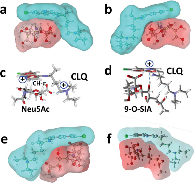
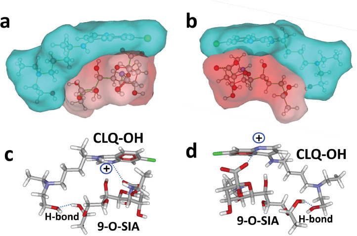
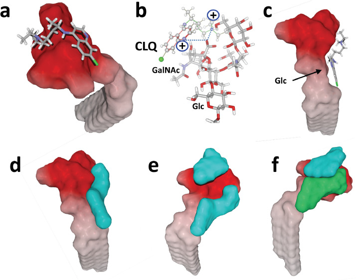
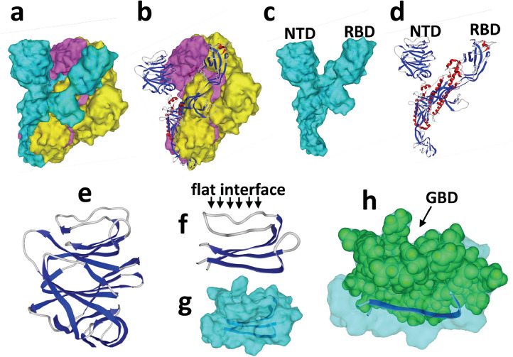
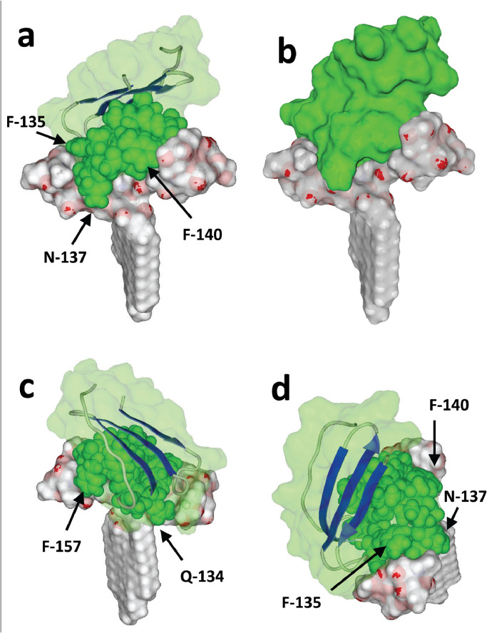
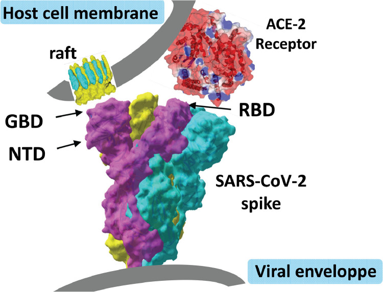
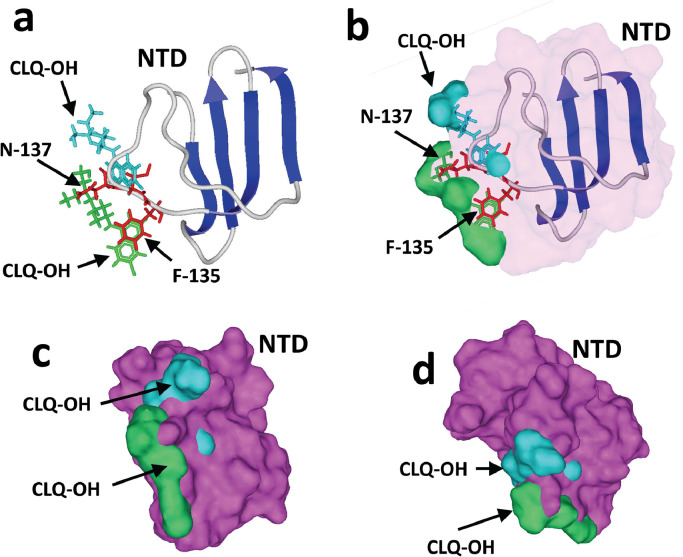
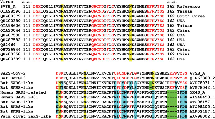
The recent emergence of the novel pathogenic SARS-coronavirus 2 (SARS-CoV-2) is responsible for a worldwide pandemic. Given the global health emergency, drug repositioning is the most reliable option to design an efficient therapy for infected patients without delay. The first step of the viral replication cycle [i.e. attachment to the surface of respiratory cells, mediated by the spike (S) viral protein] offers several potential therapeutic targets. The S protein uses the angiotension-converting enzyme-2 (ACE-2) receptor for entry, but also sialic acids linked to host cell surface gangliosides. Using a combination of structural and molecular modelling approaches, this study showed that chloroquine (CLQ), one of the drugs currently under investigation for SARS-CoV-2 treatment, binds sialic acids and gangliosides with high affinity. A new type of ganglioside-binding domain at the tip of the N-terminal domain of the SARS-CoV-2 S protein was identified. This domain (111-158), which is fully conserved among clinical isolates worldwide, may improve attachment of the virus to lipid rafts and facilitate contact with the ACE-2 receptor. This study showed that, in the presence of CLQ [or its more active derivative, hydroxychloroquine (CLQ-OH)], the viral S protein is no longer able to bind gangliosides. The identification of this new mechanism of action of CLQ and CLQ-OH supports the use of these repositioned drugs to cure patients infected with SARS-CoV-2. The in-silico approaches used in this study might also be used to assess the efficiency of a broad range of repositioned and/or innovative drug candidates before clinical evaluation.

Keywords: Chloroquine; Coronavirus; Ganglioside; Pandemic; SARS-CoV-2; Spike.


Figures

Image, graphical abstract
Graphical abstract
Fig 1
Fig. 1
Chemical structure of chloroquine (CLQ) and hydroxychloroquine (CLQ-OH). (a) CLQ. (b) CLQ-OH. (c) CLQ-OH extended conformer. (d) CLQ-OH in water. (e) Typical condensed conformer of CLQ. (f) CLQ in water. The molecules in (c–f) are shown in either tube or sphere rendering (carbon, green; nitrogen, blue; oxygen, red; hydrogen, white). In (c) and (e), the chlorine atom of CLQ and CLQ-OH is indicated by an arrow.
Fig 2
Fig. 2
Molecular modelling of chloroquine (CLQ) interaction with sialic acids. (a,b) Surface representation of the CLQ–sialic acid (Neu5Ac) complex. Two opposite views of the complex are shown. Note the geometric complementarity between the L-shape conformer of CLQ dissolved in water (in blue) and Neu5Ac (in red). (c) Neu5Ac bound to CLQ via a combination of CH-π and electrostatic interactions with one of the cationic groups of CLQ (+). (d) Molecular modelling of CLQ bound to N-acetyl-9-O-acetylneuraminic acid (9-O-SIA). From right to left, the dashed lines indicate a series of van der Waals, OH-π and electrostatic contacts with both cationic groups of CLQ (+). (e,f) Surface representations of the CLQ–9-O-SIA complex.
Fig 3
Fig. 3
Molecular modelling of hydroxychloroquine (CLQ-OH) interaction with sialic acids. (a,b) Surface representation of CLQ-OH bound to N-acetyl-9-O-acetylneuraminic acid (9-O-SIA). Two opposite views of the complex are shown. Note the geometric complementarity between CLQ-OH (in blue) and 9-O-SIA (in red). (c,d) Molecular mechanism of CLQ-OH binding to 9-O-SIA: combination of electrostatic interactions and hydrogen bonding.
Fig 4
Fig. 4
Molecular modelling simulations of chloroquine (CLQ) and hydroxychloroquine (CLQ-OH) binding to ganglioside GM1. The surface electrostatic potential of GM1 indicates a non-polar, membrane-embedded part corresponding to ceramide (white areas), and an acidic part protruding in the extracellular space corresponding to the sialic-acid-containing saccharide part (red areas). (a) CLQ bound to the tip of the carbohydrate moiety of GM1. (b) Molecular mechanism of CLQ–ganglioside interactions. (c) Molecular dynamics simulations revealed a second site of interaction. In this case, the aromatic cycles of CLQ are positioned at the ceramide–sugar junction, whereas the nitrogen atoms interact with the acidic part of the ganglioside (not illustrated). (d,e) Surface views of GM1 complexed with one (d) or two (e) CLQ molecules (both in blue), illustrating the geometric complementarity of GM1 and CLQ molecules. (f) One GM1 molecule can also accommodate two distinct CLQ-OH molecules simultaneously, after slight rearrangement allowing increased fit due to CLQ-OH/CLQ-OH interactions. To improve clarity, CLQ-OH molecules bound to GM1 are represented in two distinct colours (blue and green).
Fig 5
Fig. 5
Structural features of the SARS-CoV-2 spike (S) protein. (a) Trimeric structure (each S protein has a distinct surface colour, ‘blue’, ‘yellow’ and ‘purple’). (b) Ribbon representation of ‘blue’ S protein in the trimer (α-helix, red; β-strand, blue; coil, grey). (c) Surface structure of the ‘blue’ S protein isolated from the trimer. (d) Ribbon structure of the ‘blue’ S protein. (e) Zoom on the N-terminal domain (NTD) of the ‘blue’ S protein. (f,g) Molecular model of a minimal NTD obtained with Hyperchem [ribbon in representation in (f), surface rendering in (g)]. (h) Highlighting of the amino acid residues of the NTD that could belong to a potential ganglioside-binding domain.
Fig 6
Fig. 6
Molecular complex between the N-terminal domain (NTD) of SARS-CoV-2 spike protein and a single GM1 ganglioside. The NTD is represented in ribbons superposed with a transparent surface rendering (light green). Two symmetric views of the complex are shown (a,b). The amino acid residues Q-134 to D-138 located in the centre of the ganglioside-binding domain are represented as green spheres. The saccharide part of the ganglioside forms a landing surface for the tip of the NTD.
Fig 7
Fig. 7
Molecular complex between the N-terminal domain (NTD) of SARS-CoV-2 spike protein and a dimer of GM1. In (a), (c) and (d), the NTD is represented as in Fig. 4. In (b), the surface of the NTD is shown without any transparency. The amino acid residues Q-134 to S-162 belonging to the ganglioside-binding domain (GBD) are represented as green spheres. Compared with a single GM1 molecule, the dimer of gangliosides forms a larger attractive surface for the NTD. In the above view of (d), the anchorage of the NTD to the gangliosides is particularly obvious. As chloroquine also interacts with the saccharide part of GM1, its presence would clearly mask most of the landing surface available for the NTD, preventing attachment of the virus to the plasma membrane of host cells.
Fig 8
Fig. 8
Dual recognition of gangliosides and angiotensin-converting enzyme-2 (ACE-2) by SARS-CoV-2 spike (S) protein. The viral protein displays two distinct domains, the tips of which are available for distinct types of interactions. The receptor-binding domain binds to the ACE-2 receptor, and the N-terminal domain (NTD) binds to the ganglioside-rich domain of the plasma membrane. Lipid rafts, which are membrane domains enriched in gangliosides (in yellow) and cholesterol (in blue), provide a perfect attractive interface for adequately positioning the viral S protein at the first step of the infection process. These structural and molecular modelling studies suggest that amino acid residues 111–162 of the NTD form a functional ganglioside-binding domain, the interaction of which with lipid rafts can be efficiently prevented by chloroquine and hydroxychloroquine.
Fig 9
Fig. 9
Mechanism of action of hydroxychloroquine (CLQ-OH). The N-terminal domain (NTD) bound to GM1 was superposed onto GM1 in interaction with two CLQ-OH molecules. The models only show the NTD and both CLQ-OH molecules (not GM1, to improve clarity). (a,b) The aromatic ring of F-135 (in red), which stacks onto the glucose cycle of GM1, overlaps the aromatic CLQ-OH rings (in green) which also bind to GM1 via a CH-π stacking mechanism. The N-137 residue of the NTD interacts with the N-acetylgalactosamine residue of GM1, but this interaction cannot occur in the presence of CLQ-OH as this part of GM1 is totally masked by the drug. (c,d) Superposition of the NTD surface (in purple) with the two CLQ-OH molecules bound to GM1, illustrating the steric impossibility that prevents NTD binding to GM1 when both CLQ-OH molecules are already interacting with the ganglioside.
Fig 10
Fig. 10
Amino acid sequence alignments of the ganglioside-binding domain (GBD) of the SARS-CoV-2 spike protein. (a) Clinical SARS-CoV-2 isolates aligned with the reference sequence (6VSB_A, fragment 111–162). The amino acid residues involved in GM1 binding are indicated in red. Two asparagine residues acting as glycosylation sites are highlighted in yellow. (b) Alignments of human and animal viruses compared with SARS-CoV-2 (6VSB_A, fragment 111–162). Deletions are highlighted in green, amino acid changes in residues involved in ganglioside binding are highlighted in blue, conserved residues of the GBD are highlighted in red, and asparagine residues acting as glycosylation sites are highlighted in yellow.

Similar articles

Cited by

References

    1. Zhou P., Yang X.L., Wang X.G., Hu B., Zhang L., Zhang W. A pneumonia outbreak associated with a new coronavirus of probable bat origin. Nature. 2020;579:270–273. - PMC - PubMed
    1. Mullard A. Drug repurposing programmes get lift off. Nat Rev Drug Discov. 2012;11:505–506. - PubMed
    1. Colson P., Rolain J.M., Raoult D. Chloroquine for the 2019 novel coronavirus SARS-CoV-2. Int J Antimicrob Agents. 2020;55 - PMC - PubMed
    1. Gao J., Tian Z., Yang X. Breakthrough: chloroquine phosphate has shown apparent efficacy in treatment of COVID-19 associated pneumonia in clinical studies. Biosci Trends. 2020 Feb 19 doi: 10.5582/bst.2020.01047. [Epub ahead of print] - DOI - PubMed
    1. Savarino A., Di Trani L., Donatelli I., Cauda R., Cassone A. New insights into the antiviral effects of chloroquine. Lancet Infect Dis. 2006;6:67–69. - PMC - PubMed

MeSH terms