. 2020 Apr 1;143(4):1233-1248.
doi: 10.1093/brain/awaa068.

Medial temporal lobe connectivity and its associations with cognition in early Alzheimer's disease

Affiliations

Medial temporal lobe connectivity and its associations with cognition in early Alzheimer's disease

David Berron et al. Brain. .

Erratum in

Abstract

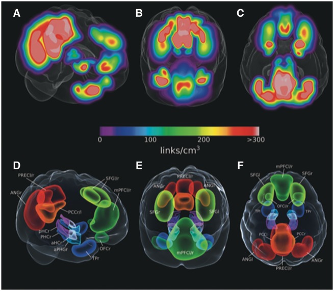
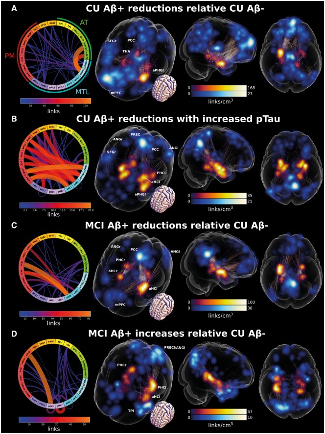
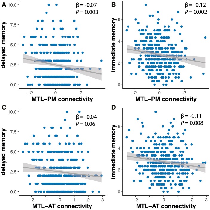
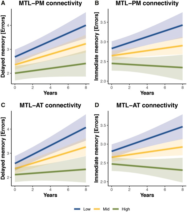
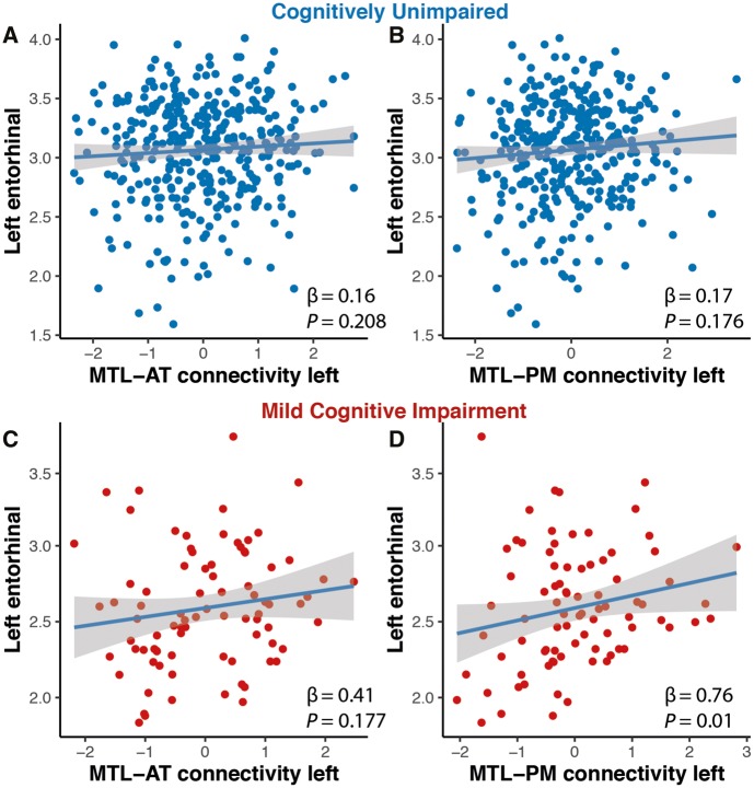
Human episodic memory critically depends on subregions of the medial temporal lobe, which are part of functional brain systems such as the anterior-temporal and the posterior-medial system. Here we analysed how Alzheimer's pathology affects functional connectivity within these systems. Data from 256 amyloid-β-negative cognitively unimpaired, 103 amyloid-β-positive cognitively unimpaired, and 83 amyloid-β-positive individuals with mild cognitive impairment were analysed. Amyloid-β and tau pathology were measured using the CSF amyloid-β42/40 ratio and phosphorylated tau, respectively. We found that amyloid-β-positive cognitively unimpaired individuals were mainly characterized by decreased functional connectivity between the medial temporal lobe and regions in the anterior-temporal system, most prominently between left perirhinal/entorhinal cortices and medial prefrontal cortex. Furthermore, correlation analysis in this group revealed decreasing functional connectivity between bilateral perirhinal/entorhinal cortices, anterior hippocampus and posterior-medial regions with increasing levels of phosphorylated tau. The amyloid-β-positive individuals with mild cognitive impairment mostly exhibited reduced connectivity between the medial temporal lobe and posterior-medial regions, predominantly between the anterior hippocampus and posterior cingulate cortex. In addition, they showed hyperconnectivity within the medial temporal lobe and its immediate proximity. Lower medial temporal-cortical functional connectivity networks resulting from the group comparisons of cognitively unimpaired individuals were associated with reduced memory performance and more rapid longitudinal memory decline as shown by linear mixed-effects regression analysis. Finally, we found that reduced medial temporal-cortical connectivity in mildly cognitively impaired individuals was related to reduced entorhinal thickness and white matter integrity of the parahippocampal cingulum and the fornix. No such relationships were found in cognitively unimpaired individuals. In conclusion, our findings show that the earliest changes in preclinical Alzheimer's disease might involve decreased connectivity within the anterior-temporal system, and early changes in connectivity might be related to memory impairment, but not to structural changes. With disease progression and increased tau pathology, medial temporal functional connectivity with posterior-medial regions seems to be increasingly impaired. In individuals with mild cognitive impairment, reduced functional connectivity is associated with structural brain changes as well as the emergence of locally increased connectivity patterns. Thus, functional connectivity between the medial temporal lobe and the anterior-temporal and posterior-medial system could serve as stage-specific functional markers in early Alzheimer's disease.

Keywords: Alzheimer’s disease; PMAT systems; fluid biomarkers; functional connectivity; medial temporal lobe.


Figures

Figure 1
Figure 1
MTL-whole brain link densities in amyloid-β− cognitively unimpaired participants and derived regions of interest from MTL-to-whole brain link densities in cognitively unimpaired participants from an additional independent dataset. (AC) MTL-to-whole brain density maps were calculated for a separate sample of cognitively unimpaired amyloid-β− participants. (DF) Cortical regions of interest were selected and delineated around coordinates with highest connectivity with the MTL. aHC = anterior hippocampus; ANG = angular gyrus; aPHG = anterior parahippocampal gyrus including entorhinal and perirhinal cortices; l = left; mPFC = medial prefrontal cortex; OFC = orbitofrontal cortex; PCC = posterior cingulate cortex; PHC = parahippocampal cortex; pHC = posterior hippocampus; PREC = precuneus; r = right; SFG = superior frontal gyrus; TP = temporal pole.
Figure 2
Figure 2
Changes in MTL-cortical functional connectivity. (A) Reduced MTL-cortical connectivity in amyloid-β+ compared to amyloid-β− cognitively unimpaired (CU) participants. (B) Decreased MTL-cortical connectivity with increasing levels of CSF p-tau in amyloid-β+ cognitively unimpaired individuals. (C) Reduced MTL-cortical connectivity in amyloid-β+ MCI patients compared to amyloid-β− cognitively unimpaired participants. (D) Increased MTL-cortical connectivity in amyloid-β+ MCI compared to amyloid-β− cognitively unimpaired participants. Significant effects are visualized as set of binary links. The end-point transparency and maximal intensity projection colour fields are proportional to the number of links converging on the voxel location. End-points in MTL regions of interest are displayed in red while end-points in cortical regions of interest are displayed in blue. The connectograms give an overview of the 3D plots, where line width and colour are proportional to the number of links between voxels constituting the respective regions of interest. Please see Supplementary Videos 1–4 for 3D visualizations. aHC = anterior hippocampus; ANG = angular gyrus; aPHG = anterior parahippocampal gyrus including entorhinal and perirhinal cortices; l = left; mPFC = medial prefrontal cortex; OFC = orbitofrontal cortex; PCC = posterior cingulate cortex; PHC = parahippocampal cortex; pHC = posterior hippocampus; PREC = precuneus; r = right; SFG = superior frontal gyrus; TP = temporal pole.
Figure 3
Figure 3
Reduced functional entropy overlaps with increased functional connectivity in a subset of amyloid-β+ MCI patients relative to amyloid-β+ cognitively unimpaired subjects. The isosurface corresponds to significantly increased entropy (calculated in 6 × 6 × 6 mm3 voxel space and convoluted with Gaussian of 6-mm FWHM to generate the visualized surface). The blue maximal intensity projection is proportional to the link density of the MCI amyloid-β+ versus cognitively unimpaired amyloid-β− functional connectivity contrast visualized.
Figure 4
Figure 4
Relationship of baseline memory scores and MTL-cortical connectivity estimates. Delayed and immediate recall were associated with MTL-PM functional connectivity (A and B) while only immediate recall was related to MTL-AT functional connectivity (C and D).
Figure 5
Figure 5
Relationship of MTL-cortical connectivity estimates and memory decline. Plots of estimated curves for three groups with different baseline functional connectivity (low, mid, high) and memory outcomes over time. Cognitively unimpaired (CU) individuals are subdivided in tertiles according to their baseline MTL-PM and MTL-AT functional connectivity. The lowest tertile is shown in blue, the middle in yellow and the highest in green. Estimation of curves included all participants regardless of length of follow-up time. Lower baseline MTL-PM connectivity (A and B) and lower baseline MTL-AT functional connectivity (C and D) were associated with steeper decline rates in immediate and delayed memory (increase in number of errors).
Figure 6
Figure 6
Relationship of white matter tract integrity and system-specific functional connectivity estimates. In MCI patients, baseline MTL-PM functional connectivity was associated with fractional anisotropy of the left parahippocampal cingulum (C). Baseline MTL-AT connectivity was associated with fractional anisotropy of the fornix (D). There were no such relationships for cognitively unimpaired individuals (A and B). FA = fractional anisotropy; PC = parahippocampal cingulum.
Figure 7
Figure 7
Relationship of entorhinal thickness and system-specific functional connectivity estimates. In MCI patients, baseline MTL-PM (D) but not MTL-AT functional connectivity was associated with thickness of the left entorhinal cortex (C). There were no such relationships for cognitively unimpaired individuals (A and B).

Similar articles

Cited by

References

    1. Avants BB, Tustison NJ, Stauffer M, Song G, Wu B, Gee JC.. The Insight ToolKit image registration framework. Front Neuroinform 2014; 8: 44. - PMC - PubMed
    1. Badhwar A, Tam A, Dansereau C, Orban P, Hoffstaedter F, Bellec P.. Resting-state network dysfunction in Alzheimer’s disease: a systematic review and meta-analysis. Alzheimer’s & Dementia: diagnosis. Alzheimers Dement 2017; 8: 73–85. - PMC - PubMed
    1. Behzadi Y, Restom K, Liau J, Liu TT.. A component based noise correction method (CompCor) for BOLD and perfusion based fMRI. Neuroimage 2007; 37: 90–101. - PMC - PubMed
    1. Berron D, Neumann K, Maass A, Schütze H, Fliessbach K, Kiven V, et al.Age-related functional changes in domain-specific medial temporal lobe pathways. Neurobiol Aging 2018; 65: 86–97. - PubMed
    1. Berron D, Vieweg P, Hochkeppler A, Pluta J, Ding S-L, Maass A, et al.A protocol for manual segmentation of medial temporal lobe subregions in 7 Tesla MRI. Neuroimage Clin 2017; 15: 466–82. - PMC - PubMed

Publication types