

Applications of Carbon Nanotubes in Bone Regenerative Medicine
- PMID: 32252244
- PMCID: PMC7221610
- DOI: 10.3390/nano10040659
Applications of Carbon Nanotubes in Bone Regenerative Medicine
Abstract
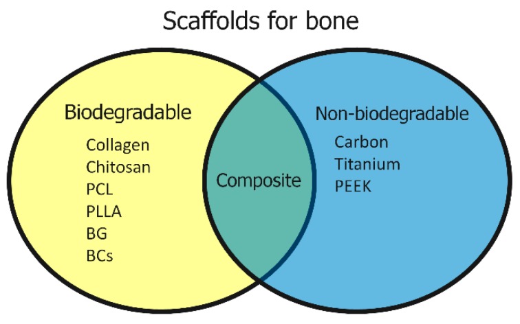
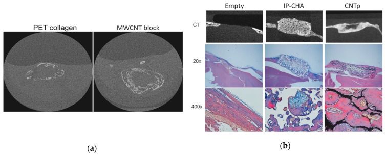
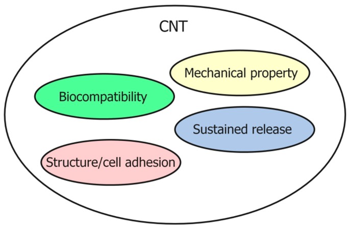
Scaffolds are essential for bone regeneration due to their ability to maintain a sustained release of growth factors and to provide a place where cells that form new bone can enter and proliferate. In recent years, scaffolds made of various materials have been developed and evaluated. Functionally effective scaffolds require excellent cell affinity, chemical properties, mechanical properties, and safety. Carbon nanotubes (CNTs) are fibrous nanoparticles with a nano-size diameter and have excellent strength and chemical stability. In the industrial field, they are used as fillers to improve the performance of materials. Because of their excellent physicochemical properties, CNTs are studied for their promising clinical applications as biomaterials. In this review article, we focused on the results of our research on CNT scaffolds for bone regeneration, introduced the promising properties of scaffolds for bone regeneration, and described the potential of CNT scaffolds.
Keywords: bone defects; bone regeneration; carbon nanotubes; scaffolds.
Conflict of interest statement
The authors declare no conflict of interest.
Figures
Similar articles
-
In situ hybridization of carbon nanotubes with bacterial cellulose for three-dimensional hybrid bioscaffolds.Biomaterials. 2015 Jul;58:93-102. doi: 10.1016/j.biomaterials.2015.04.027. Epub 2015 May 11. Biomaterials. 2015. PMID: 25941786
-
Applications of Carbon Nanotubes in Bone Tissue Regeneration and Engineering: Superiority, Concerns, Current Advancements, and Prospects.Nanomaterials (Basel). 2019 Oct 22;9(10):1501. doi: 10.3390/nano9101501. Nanomaterials (Basel). 2019. PMID: 31652533 Free PMC article. Review.
-
Carbon nanotube-based biomaterials for orthopaedic applications.J Mater Chem B. 2020 Oct 21;8(40):9227-9238. doi: 10.1039/d0tb01440k. J Mater Chem B. 2020. PMID: 32935730 Review.
-
Recent Progress in Carbon Nanotube Polymer Composites in Tissue Engineering and Regeneration.Int J Mol Sci. 2020 Sep 3;21(17):6440. doi: 10.3390/ijms21176440. Int J Mol Sci. 2020. PMID: 32899409 Free PMC article. Review.
-
Carbon nanotubes for biomaterials in contact with bone.Curr Med Chem. 2008;15(5):523-7. doi: 10.2174/092986708783503140. Curr Med Chem. 2008. PMID: 18289008 Review.
Cited by
-
Nanoparticles and Nanomaterials: A Review from the Standpoint of Pharmacy and Medicine.Pharmaceutics. 2025 May 16;17(5):655. doi: 10.3390/pharmaceutics17050655. Pharmaceutics. 2025. PMID: 40430945 Free PMC article. Review.
-
A Structural Analysis of Proteinaceous Nanotube Cavities and Their Applications in Nanotechnology.Nanomaterials (Basel). 2022 Nov 20;12(22):4080. doi: 10.3390/nano12224080. Nanomaterials (Basel). 2022. PMID: 36432365 Free PMC article. Review.
-
Frame Coating of Single-Walled Carbon Nanotubes in Collagen on PET Fibers for Artificial Joint Ligaments.Int J Mol Sci. 2020 Aug 26;21(17):6163. doi: 10.3390/ijms21176163. Int J Mol Sci. 2020. PMID: 32859107 Free PMC article.
-
Current Methods in the Study of Nanomaterials for Bone Regeneration.Nanomaterials (Basel). 2022 Apr 2;12(7):1195. doi: 10.3390/nano12071195. Nanomaterials (Basel). 2022. PMID: 35407313 Free PMC article. Review.
-
A non-invasive smart scaffold for bone repair and monitoring.Bioact Mater. 2022 May 6;19:499-510. doi: 10.1016/j.bioactmat.2022.04.034. eCollection 2023 Jan. Bioact Mater. 2022. PMID: 35600976 Free PMC article.
References
Publication types
LinkOut - more resources
Full Text Sources
