. 2020 Apr 2;25(7):1630.
doi: 10.3390/molecules25071630.

Walnut Oil Prevents Scopolamine-Induced Memory Dysfunction in a Mouse Model

Affiliations

Walnut Oil Prevents Scopolamine-Induced Memory Dysfunction in a Mouse Model

Jianqiao Liao et al. Molecules. .

Abstract

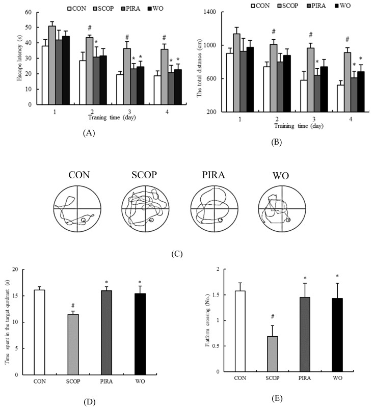
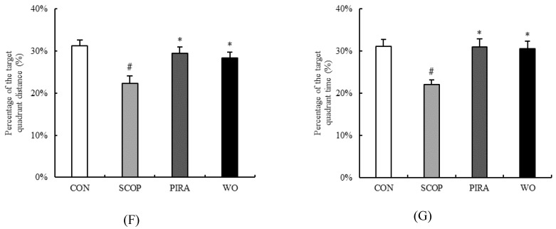
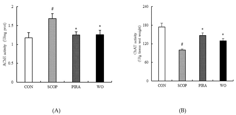
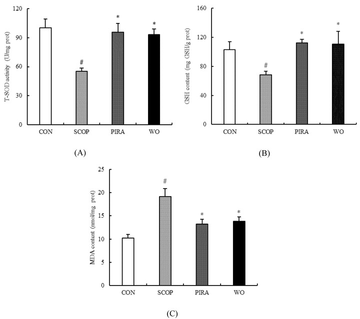
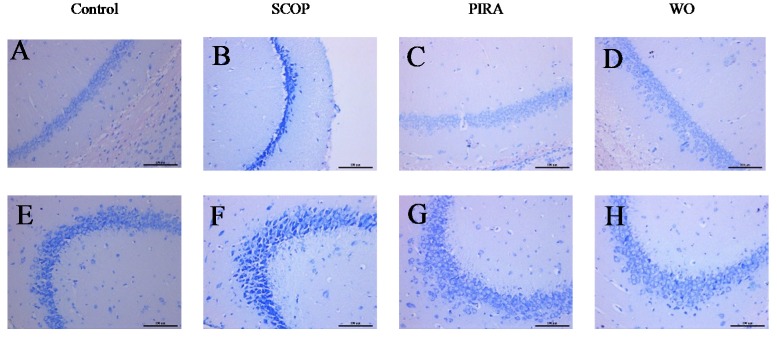
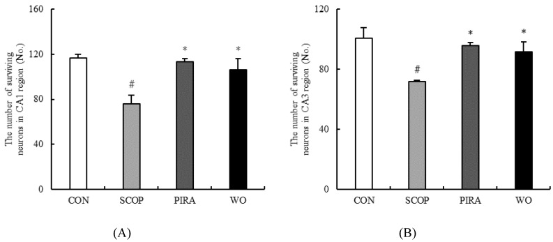
For thousands of years, it has been widely believed that walnut is a kind of nut that has benefits for the human body. Walnut oil, accounting for about 70% of walnut, mainly consists of polyunsaturated fatty acids. To investigate the effect of walnut oil on memory impairment in mice, scopolamine (3 mg/kg body weight/d) was used to establish the animal model during Morris Water Maze (MWM) tests. Walnut oil was administrated orally at 10 mL/kg body weight/d for 8 consecutive weeks. The results showed that walnut oil treatment ameliorated the behavior of the memory-impaired mice in the MWM test. Additionally, walnut oil obviously inhibited acetylcholinesterase activity (1.26 ± 0.12 U/mg prot) (p = 0.013) and increased choline acetyltransferase activity (129.75 ± 6.76 U/mg tissue wet weight) in the brains of scopolamine-treated mice (p = 0.024), suggesting that walnut oil could prevent cholinergic function damage in mice brains. Furthermore, walnut oil remarkably prevented the decrease in total superoxide dismutase activity (93.30 ± 5.50 U/mg prot) (p = 0.006) and glutathione content (110.45 ± 17.70 mg/g prot) (p = 0.047) and the increase of malondialdehyde content (13.79 ± 0.96 nmol/mg prot) (p = 0.001) in the brain of scopolamine-treated mice, indicating that walnut oil could inhibit oxidative stress in the brain of mice. Furthermore, walnut oil prevented histological changes of neurons in hippocampal CA1 and CA3 regions induced by scopolamine. These findings indicate that walnut oil could prevent memory impairment in mice, which might be a potential way for the prevention of memory dysfunctions.

Keywords: cholinergic system; memory impairment; oxidative stress; scopolamine-induced; walnut oil.


Conflict of interest statement

The authors declare that they have no conflicts of interest.

Figures

Figure 1
Figure 1
Effects of walnut oil on escape latency (A) and total swimming distance (B) from day1 to day 4, and number of platform crossing (C), time spent in the target quadrant (D), percentage of the target quadrant distance (E), percentage of the target quadrant time (F) and represent tracks of mice (G) on day 5 during the Morris water maze test. Data are shown as mean ± SEM. # p < 0.05 versus the control group. *p < 0.05 versus the SCOP-treated group.
Figure 1
Figure 1
Effects of walnut oil on escape latency (A) and total swimming distance (B) from day1 to day 4, and number of platform crossing (C), time spent in the target quadrant (D), percentage of the target quadrant distance (E), percentage of the target quadrant time (F) and represent tracks of mice (G) on day 5 during the Morris water maze test. Data are shown as mean ± SEM. # p < 0.05 versus the control group. *p < 0.05 versus the SCOP-treated group.
Figure 2
Figure 2
Effects of walnut oil on the activity of AChE (A) and ChAT (B) in brains of mice. Data are shown as mean ± SEM. # p < 0.05 versus the control group. *p < 0.05 versus the SCOP-treated group.
Figure 3
Figure 3
Effects of walnut oil on the activity of T-SOD (A), the contents of GSH (B) and MDA (C) in brains of mice. Data are shown as mean ± SEM. #p <0.05 versus the control group. *p <0.05 versus the SCOP-treated group.
Figure 4
Figure 4
Effects of walnut oil on the result of Nissl staining of hippocampal CA1 (AD) and CA3 (EH) regions of brains of mice. Histological sections of the brain tissue showing neurological lesions (AH). Scale bar is 100 µm.
Figure 5
Figure 5
Effect of walnut on the number of surviving neurons in hippocampal CA1 (A) and CA3 (B) regions. Data are shown as mean ± SEM. # p <0.05 versus the control group. *p <0.05 versus the SCOP-treated group.

References

    1. Francis P.T., Palmer A.M., Snape M., Wilcock G.K. The cholinergic hypothesis of Alzheimer’s disease: A review of progress—Reply. J. Neurol. Neurosur. Psychiatry. 1999;67:558. doi: 10.1136/jnnp.66.2.137. - DOI - PMC - PubMed
    1. Anand R., Gill K.D., Mahdi A.A. Therapeutics of Alzheimer’s disease: Past, present and future. Neuropharmacology. 2014;76:27–50. doi: 10.1016/j.neuropharm.2013.07.004. - DOI - PubMed
    1. Schliebs R. Basal Forebrain Cholinergic Dysfunction in Alzheimer’s Disease—Interrelationship with β-amyloid, Inflammation and Neurotrophin Signaling. Neurochem. Res. 2005;30:895–908. doi: 10.1007/s11064-005-6962-9. - DOI - PubMed
    1. Sultana R., Butterfield D.A. Role of Oxidative Stress in the Progression of Alzheimer’s Disease. J. Alzheimers Dis. 2010;19:341–353. doi: 10.3233/JAD-2010-1222. - DOI - PubMed
    1. Ulep M.G., Saraon S.K., McLea S. Alzheimer Disease. J. Nurse Pr. 2018;14:129–135. doi: 10.1016/j.nurpra.2017.10.014. - DOI

MeSH terms