. 2020 Apr 2;9(4):858.
doi: 10.3390/cells9040858.

Modulation of STAT3 Signaling, Cell Redox Defenses and Cell Cycle Checkpoints by β-Caryophyllene in Cholangiocarcinoma Cells: Possible Mechanisms Accounting for Doxorubicin Chemosensitization and Chemoprevention

Affiliations

Modulation of STAT3 Signaling, Cell Redox Defenses and Cell Cycle Checkpoints by β-Caryophyllene in Cholangiocarcinoma Cells: Possible Mechanisms Accounting for Doxorubicin Chemosensitization and Chemoprevention

Antonella Di Sotto et al. Cells. .

Abstract

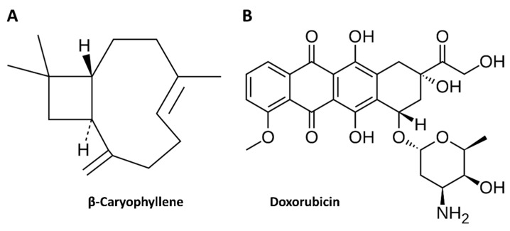
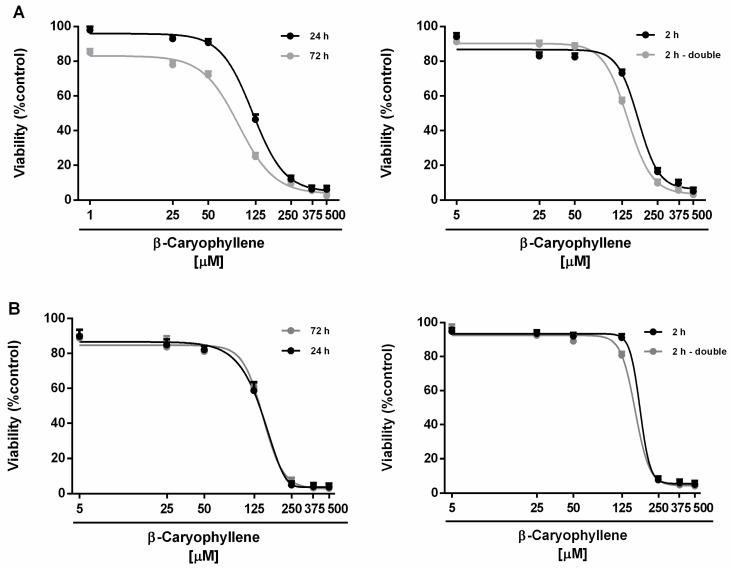
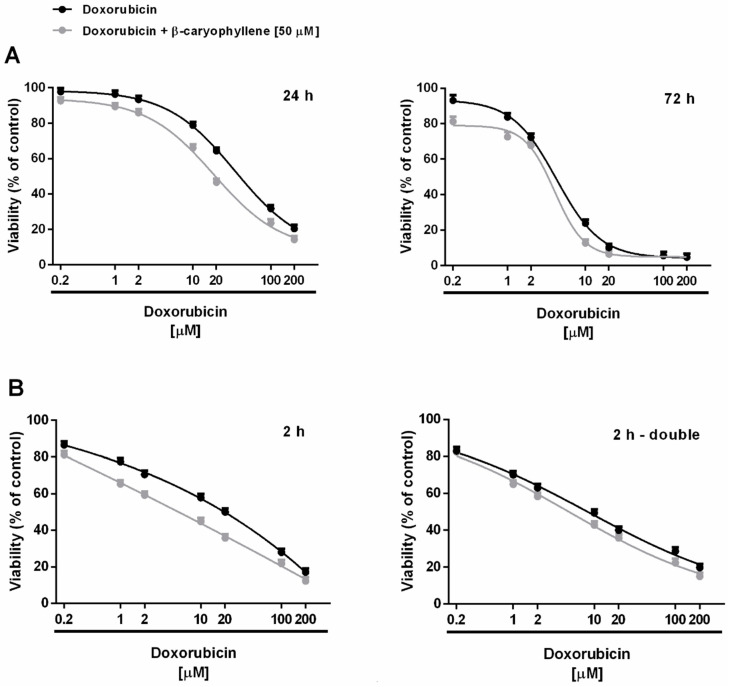
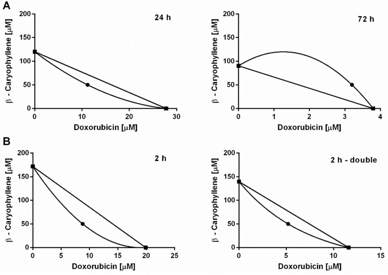
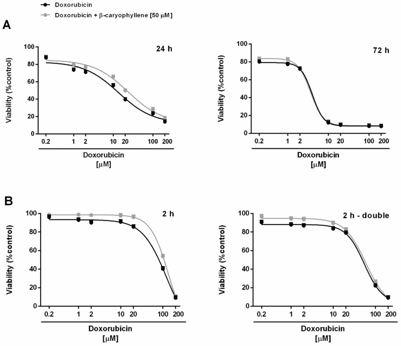
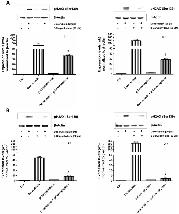
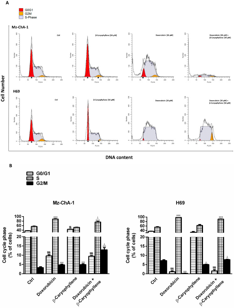
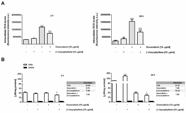
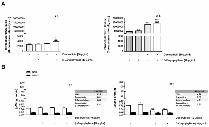
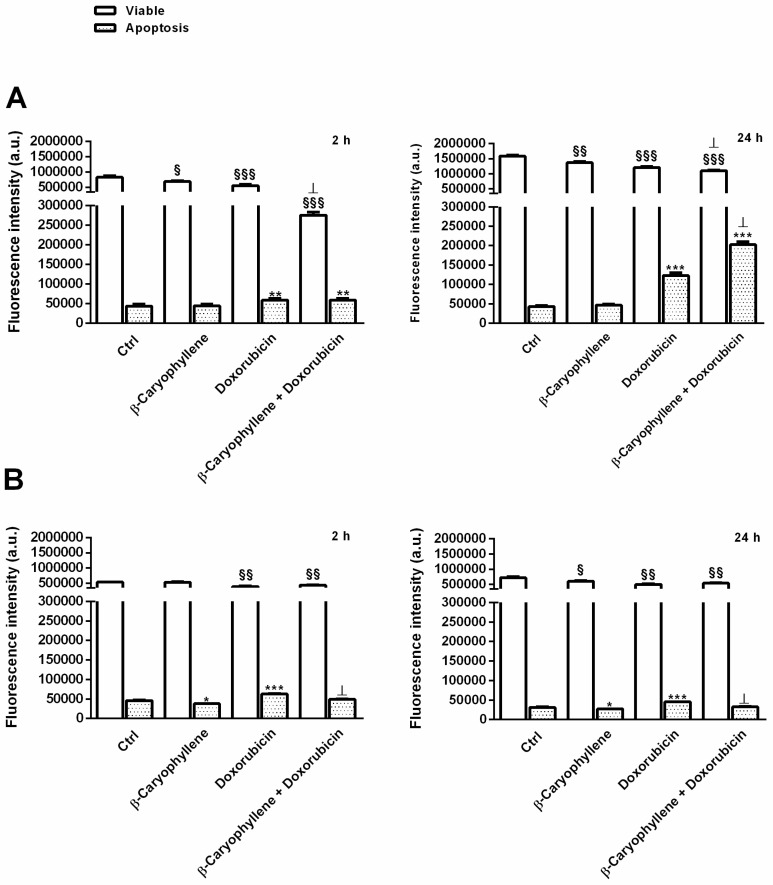
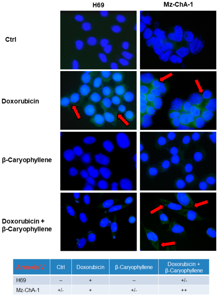
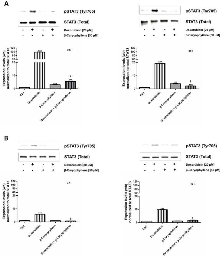
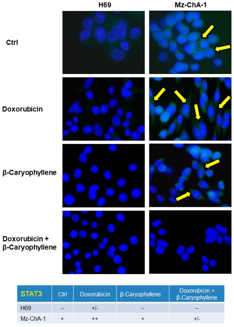
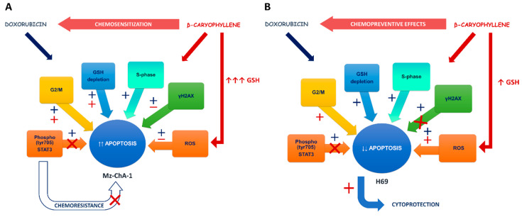
Cholangiocarcinoma (CCA) is an aggressive group of biliary tract cancers, characterized by late diagnosis, low effective chemotherapies, multidrug resistance, and poor outcomes. In the attempt to identify new therapeutic strategies for CCA, we studied the antiproliferative activity of a combination between doxorubicin and the natural sesquiterpene β-caryophyllene in cholangiocarcinoma Mz-ChA-1 cells and nonmalignant H69 cholangiocytes, under both long-term and metronomic schedules. The modulation of STAT3 signaling, oxidative stress, DNA damage response, cell cycle progression and apoptosis was investigated as possible mechanisms of action. β-caryophyllene was able to synergize the cytotoxicity of low dose doxorubicin in Mz-ChA-1 cells, while producing cytoprotective effects in H69 cholangiocytes, mainly after a long-term exposure of 24 h. The mechanistic analysis highlighted that the sesquiterpene induced a cell cycle arrest in G2/M phase along with the doxorubicin-induced accumulation in S phase, reduced the γH2AX and GSH levels without affecting GSSG. ROS amount was partly lowered by the combination in Mz-ChA-1 cells, while increased in H69 cells. A lowered expression of doxorubicin-induced STAT3 activation was found in the presence of β-caryophyllene in both cancer and normal cholangiocytes. These networking effects resulted in an increased apoptosis rate in Mz-ChA-1 cells, despite a lowering in H69 cholangiocytes. This evidence highlighted a possible role of STAT3 as a final effector of a complex network regulated by β-caryophyllene, which leads to an enhanced doxorubicin-sensitivity of cholangiocarcinoma cells and a lowered chemotherapy toxicity in nonmalignant cholangiocytes, thus strengthening the interest for this natural sesquiterpene as a dual-acting chemosensitizing and chemopreventive agent.

Keywords: GSH depletion; H2AX phosphorylation; STAT3 signaling; apoptosis; caryophyllane sesquiterpenes; cell cycle checkpoint; chemoprevention; cholangiocytes; genoprotective effects; liver cancer; metronomic schedule.


Conflict of interest statement

The authors declare no conflict of interest.

Figures

Figure 1
Figure 1
Molecular structure of the natural sesquiterpene β-caryophyllene (A) and the anticancer drug doxorubicin (B).
Figure 2
Figure 2
Cytotoxicity of β-caryophyllene in Mz-ChA-1 cholangiocarcinoma cells (A) and H69 cholangiocytes (B) under both the single long-term exposures of 24 h and 72 h and the metronomic schedule. In the last protocol, the cells were subjected to a single and/or double repeated short exposure of 2 h followed by a recovery time of 72 h. Data are expressed as mean ± SE (standard error) of at least two experiments in which each treatment was tested at least in triplicate (n = 6).
Figure 3
Figure 3
Cytotoxicity of doxorubicin and its combination with β-caryophyllene in Mz-ChA-1 cholangiocarcinoma cells. (A) Single long-term exposures of 24 h and 72 h. (B) Metronomic schedule: the cells were subjected to a single and/or double repeated short exposure of 2 h followed by a recovery time of 72 h. Data are expressed as mean ± SE (standard error) of at least two experiments in which each treatment was tested at least in triplicate (n = 6).
Figure 4
Figure 4
Isobolographic analysis of the cytotoxicity produced by doxorubicin in combination with β-caryophyllene at the chemosensitizing concentrations of 50 µM in Mz-ChA-1 cholangiocarcinoma cells. (A) Long-term exposure of 24 h and 72 h. (B) Single and double repeated metronomic exposure of 2 h.
Figure 5
Figure 5
Cytotoxicity of doxorubicin and its combination with β-caryophyllene in H69 noncancerous cholangiocytes. (A) Single long-term exposures of 24 h and 72 h. (B) Metronomic schedule: the cells were subjected to a single and/or double repeated short exposure of 2 h followed by a recovery time of 72 h. Data are expressed as mean ± SE (standard error) of at least two experiments in which each treatment was tested at least in triplicate (n = 6).
Figure 6
Figure 6
Effect of the natural sesquiterpene β-caryophyllene (50 µM), doxorubicin (20 µM) and their combination compared the control on the levels of phosphorylated H2AX at Ser139 residue (γH2AX) in Mz-ChA-1 cholangiocarcinoma cells (A) and H69 noncancerous cholangiocytes (B). The cells were treated for 2 h and 24 h, then the pellets were harvested for the western blotting analysis. For each experimental condition, the densitometric bar graph (data expressed as mean ± standard error) obtained from at least two independent replicates and a representative western blotting image, showing the expression levels of γH2AX and β-actin used as protein loading control, were displayed. ***p < 0.001 (one-way ANOVA followed by Dunnett’s multiple comparison post-test), significantly higher than the vehicle control (basal level). §p < 0.001 (t-Student test), significantly lower than doxorubicin.
Figure 7
Figure 7
Effect of the natural sesquiterpene β-caryophyllene (50 µM), doxorubicin (20 µM) and their combination compared the control on the cell cycle progression in Mz-ChA-1 cholangiocarcinoma cells and H69 noncancerous cholangiocytes. The cells were treated for 24 h, then the pellets were harvested, fixed with 70% ethanol and stained with propidium iodide for the cytofluorimetric analysis. (A) Representative histograms showing the percentages of cells in different cell cycle phases after treatments in Mz-ChA-1 and H69 cells. (B) Bar graph analysis obtained from at least two independent replicates (data expressed as mean ± standard error). §§p < 0.01 and §§§p < 0.001 (one-way ANOVA followed by Dunnett’s multiple comparison post-test) denote a significant difference of G0/G1 phase in the treatments compared the control. ***p < 0.001 (one-way ANOVA followed by Dunnett’s multiple comparison post-test) denotes a significant difference of S phase in the treatments compared the control.). °p < 0.05 and °°°p < 0.001 (one-way ANOVA followed by Dunnett’s multiple comparison post-test) denotes a significant difference of G2/M phase in the treatments compared the control. p < 0.001 (t-Student test) denotes a significant difference with respect to doxorubicin.
Figure 8
Figure 8
Effect of the natural sesquiterpene β-caryophyllene (50 µM), doxorubicin (20 µM) and their combination compared the control on the intracellular oxidative stress in Mz-ChA-1 cholangiocarcinoma cells. The cells were treated for 2 h and 24 h, then the pellets were harvested for the subsequent analysis. (A) Bar graphs representing the levels of reactive oxygen species (ROS) as detected by the 2,7-dichlorofluorescein diacetate (DCFH-DA) assay. Data are expressed as mean ± standard error of at least two independent replicates. (B) Bar graphs representing the levels of GSH (reduced glutathione) and GSSG (oxidized glutathione) as revealed by HPLC analysis. Data are expressed as mean ± standard error of at least two independent replicates. °p < 0.05, °°p < 0.01 and °°°p < 0.001 (one-way ANOVA followed by Dunnett’s multiple comparison post-test) denote a significant difference of the treatments compared the control. *p < 0.05 and ***p < 0.001 (t-Student test) denote a significant difference with respect to doxorubicin.
Figure 9
Figure 9
Effect of the natural sesquiterpene β-caryophyllene (50 µM), doxorubicin (20 µM) and their combination compared the control on the intracellular oxidative stress in H69 noncancerous cholangiocytes. The cells were treated for 2 h and 24 h, then the pellets were harvested for the subsequent analysis. (A) Bar graphs representing the levels of reactive oxygen species (ROS) as detected by the 2,7-dichlorofluorescein diacetate (DCFH-DA) assay. Data are expressed as mean ± standard error of at least two independent replicates. (B) Bar graphs representing the levels of GSH (reduced glutathione) and GSSG (oxidized glutathione) as revealed by HPLC analysis. Data are expressed as mean ± standard error of at least two independent replicates. °p < 0.05, °°p < 0.01 and °°°p < 0.001 (one-way ANOVA followed by Dunnett’s multiple comparison post-test) denote a significant difference of the treatments compared the control. *p < 0.05 and ***p < 0.001 (t-Student test) denote a significant difference with respect to doxorubicin.
Figure 10
Figure 10
Bar graphs (data expressed as mean ± standard error at least two independent replicates) of apoptosis induced by the natural sesquiterpene β-caryophyllene (50 µM), doxorubicin (20 µM) and their combination compared the control in Mz-ChA-1 cholangiocarcinoma cells (A) and H69 noncancerous cholangiocytes (B). The cells were treated for 2 h and 24 h, then the pellets were harvested and stained with Annexin-V-Cy3 and carboxyfluorescein diacetate (CFDA) for the cytofluorimetric analysis of apoptotic and viable cells (FL-2 for annexin and FL-1 for CF; BD AccuriTM C6 flow cytometer). Cells undergoing apoptosis were sorted by their typical forward- and side scatter (FSC-SSC) pattern, i.e., increased SSC and decreased FSC, respect to the viable cells. §p < 0.05, §§p < 0.01 and §§§p < 0.001 (one-way ANOVA followed by Dunnett’s multiple comparison post-test) denote a significant difference in the viable cells of the treatments compared the control. *p < 0.05, **p < 0.01 and ***p < 0.001 (one-way ANOVA followed by Dunnett’s multiple comparison post-test) denote a significant difference in the apoptotic cells of the treatments compared the control. p < 0.01 (t-Student test) denotes a significant difference with respect to doxorubicin.
Figure 11
Figure 11
Representative immunofluorescence (IF) images and semiquantitative analysis of the apoptosis induced by the natural sesquiterpene β-caryophyllene (50 µM), doxorubicin (20 µM) and their combination compared the control in Mz-ChA-1 cholangiocarcinoma cells and H69 noncancerous cholangiocytes. After treatments of 24 h, the cells were stained with Annexin-V-FITC to assess the apoptotic rate, as shown by the red arrows. The semiquantitative analysis has been carried out (four fields for each treatment) applying a previous published grading system [64]: 0%–5% = negative; 6%–10% = +/−; 11%–30% = +; 31%–60% = ++; > 61% = +++.
Figure 12
Figure 12
Effect of the natural sesquiterpene β-caryophyllene (50 µM), doxorubicin (20 µM) and their combination compared the control on the expression levels of phosphorylated STAT3 on tyrosine 705 residue in Mz-ChA-1 cholangiocarcinoma cells (A) and H69 noncancerous cholangiocytes (B). The cells were treated for 2 h and 24 h, then the pellets were harvested for the western blotting analysis. For each experimental condition, a representative western blotting image, showing the expression levels of the phospho(Tyr705) STAT3 and total STAT3 used as protein loading control and a densitometric bar graph analysis (data expressed as mean ± standard error) obtained from at least two independent replicates, were displayed. *** p < 0.001 (one-way ANOVA followed by Dunnett’s multiple comparison post-test) denotes a significant increase compared the control. § p < 0.001 (t-Student test) denotes a significant reduction with respect to doxorubicin.
Figure 13
Figure 13
Representative immunofluorescence (IF) images and semiquantitative analysis of the phosphorylated STAT3 on tyrosine 705 residue induced by the natural sesquiterpene β-caryophyllene (50 µM), doxorubicin (20 µM) and their combination compared the control in Mz-ChA-1 cholangiocarcinoma cells and H69 noncancerous cholangiocytes. After treatments of 24 h, the cells were fixed then stained with a specific anti-phospho(Tyr705)-STAT3 primary antibody to assess the protein phosphorylation rate, as shown by the yellow arrows. The semiquantitative analysis has been carried out (four fields for each treatment) applying a previous published grading system [64]: 0%–5% = negative; 6%–10% = +/−; 11%–30% = +; 31%–60% = ++; > 61% = +++.
Figure 14
Figure 14
Scheme of the possible network involved in the chemosensitizing and chemopreventive effects of β-caryophyllene towards doxorubicin in Mz-ChA-1 cholangiocarcinoma cells (A) and in H69 noncancerous cholangiocytes (B). Doxorubicin-induced oxidative stress, measured by the intracellular levels of reactive oxygen species (ROS), was partly reduced in combination with β-caryophyllene in Mz-ChA-1 cells, whereas GSH defenses, markedly upregulated by the sesquiterpene alone, were drastically reduced by both doxorubicin and its combination with β-caryophyllene, without affecting GSSG amount. Conversely, in H69 cholangiocytes, despite a GSH depletion similar to doxorubicin and not correlated with an increase in GSSG, the combined treatment of doxorubicin and β-caryophyllene enhanced ROS levels with respect to both control and the anticancer drug alone, despite a slight increase of GSH by the only β-caryophyllene. γH2AX, a biomarker of doxorubicin-induced DNA-damage, resulted partly lowered by the presence of β-caryophyllene in cholangiocarcinoma cells, with an almost complete inhibition in normal cholangiocytes. The increased G2/M checkpoint phase by β-caryophyllene could allow to repair the doxorubicin-damaged DNA in H69 cholangiocytes. In both cell lines, the phosphorylation of STAT3 at tyrosine 705 site, induced by the anticancer drug, resulted markedly lowered by the natural sesquiterpene. These networking effects result in an increased apoptotic fate in cholangiocarcinoma Mz-ChA-1 cells, despite an inhibition apoptosis in H69 noncancerous cholangiocytes.

Similar articles

Cited by

References

    1. Razumilava N., Gores G.J. Cholangiocarcinoma. Lancet. 2014;383:2168–2179. doi: 10.1016/S0140-6736(13)61903-0. - DOI - PMC - PubMed
    1. Fava G., DeMorrow S., Gaudio E., Franchitto A., Onori P., Carpino G., Glaser S., Francis H., Coufal M., Marucci L., et al. Endothelin inhibits cholangiocarcinoma growth by a decrease in the vascular endothelial growth factor expression. Liver Int. 2009;29:1031–1042. doi: 10.1111/j.1478-3231.2009.01997.x. - DOI - PMC - PubMed
    1. Rizvi S., Khan S.A., Hallemeier C.L., Kelley R.K., Gores G.J. Cholangiocarcinoma—Evolving concepts and therapeutic strategies. Nat. Rev. Clin. Oncol. 2017;15:95–111. doi: 10.1038/nrclinonc.2017.157. - DOI - PMC - PubMed
    1. Adeva J., Sangro B., Salati M., Edeline J., La Casta A., Bittoni A., Berardi R., Bruix J., Valle J.W., Berardi R. Medical treatment for cholangiocarcinoma. Liver Int. 2019;39:123–142. doi: 10.1111/liv.14100. - DOI - PubMed
    1. Lozano E., Macias R.I., Monte M.J., Asensio M., Del Carmen S., Sanchez-Vicente L., Alonso-Peña M., Al-Abdulla R., Munoz-Garrido P., Satriano L., et al. Causes of hOCT1-Dependent Cholangiocarcinoma Resistance to Sorafenib and Sensitization by Tumor-Selective Gene Therapy. Hepatology. 2019;70:1246–1261. doi: 10.1002/hep.30656. - DOI - PubMed

Publication types

MeSH terms

LinkOut - more resources