Review
. 2020 Apr 2;9(4):862.
doi: 10.3390/cells9040862.

New Insights from IGF-IR Stimulating Activity Analyses: Pathological Considerations

Affiliations
Review

New Insights from IGF-IR Stimulating Activity Analyses: Pathological Considerations

Joseph A M J L Janssen. Cells. .

Abstract

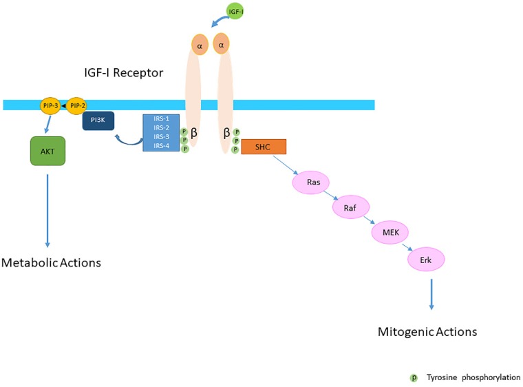
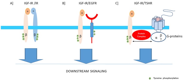
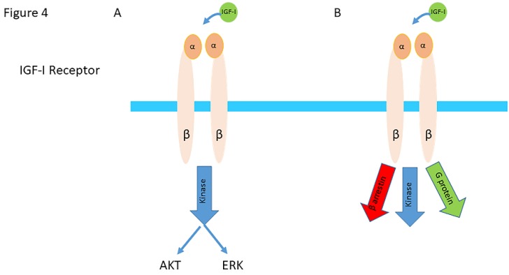
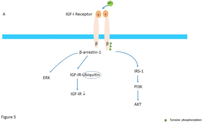
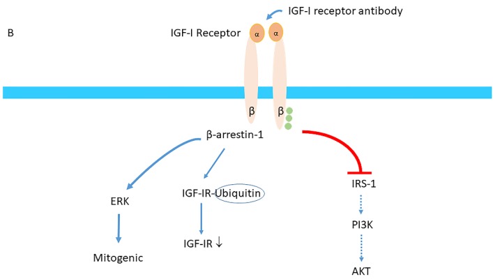
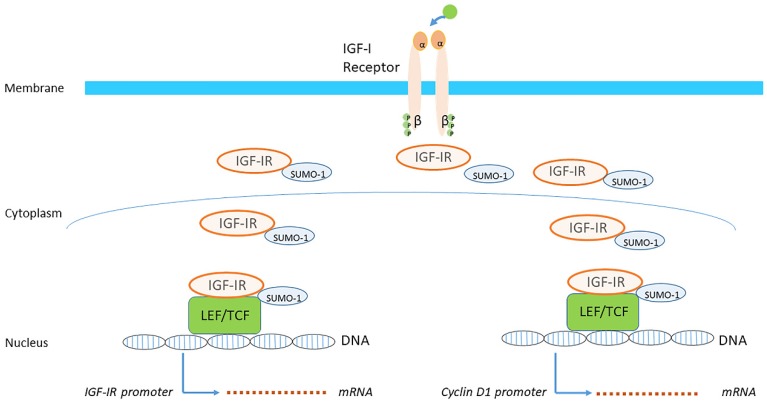
Insulin-like growth factor-I (IGF-I) and insulin-like growth factor-II (IGF-II) play a crucial factor in the growth, differentiation and survival of cells in health and disease. IGF-I and IGF-II primarily activate the IGF-I receptor (IGF-IR), which is present on the cell surface. Activation of the IGF-IR stimulates multiple pathways which finally results in multiple biological effects in a variety of tissues and cells. In addition, activation of the IGF-IR has been found to be essential for the growth of cancers. The conventional view in the past was that the IGF-IR was exclusively a tyrosine kinase receptor and that phosphorylation of tyrosine residues, after binding of IGF-I to the IGF-IR, started a cascade of post-receptor events. Recent research has shown that this view was too simplistic. It has been found that the IGF-IR also has kinase-independent functions and may even emit signals in the unoccupied state through some yet-to-be-defined non-canonical pathways. The IGF-IR may further form hybrids with the insulin receptors but also with receptor tyrosine kinases (RTKs) outside the insulin-IGF system. In addition, the IGF-IR has extensive cross-talk with many other receptor tyrosine kinases and their downstream effectors. Moreover, there is now emerging evidence that the IGF-IR utilizes parts of the G-protein coupled receptor (GPCR) pathways: the IGF-IR can be considered as a functional RTK/GPCR hybrid, which integrates the kinase signaling with some IGF-IR mediated canonical GPCR characteristics. Like the classical GPCRs the IGF-IR can also show homologous and heterologous desensitization. Recently, it has been found that after activation by a ligand, the IGF-IR may be translocated into the nucleus and function as a transcriptional cofactor. Thus, in recent years, it has become clear that the IGF-IR signaling pathways are much more complex than first thought. Therefore a big challenge for the (near) future will be how all the new knowledge about IGF-IR signaling can be translated into the clinical practice and improve diagnosis and treatment of diseases.

Keywords: G-proteins; GPCRs; IGF-I; IGF-II; IGF-IR; IRs; functional RTK/GPCR hybrid; hybrids; insulin; nuclear translocation; phosphorylation; tyrosine kinase receptor; β-arrestins.


Conflict of interest statement

The author declares no conflict of interest.

Figures

Figure 1
Figure 1
The Insulin-like Growth Factor-I (IGF-IR) is a transmembrane protein composed of two alpha (α) and two beta (β) subunits. The conventional view was that the IGF-IR was exclusively a tyrosine kinase receptor and that the binding of IGF-I to the IGF-IR started the intracellular downstream signal cascade. In this model IGF-I or IGF-II binding to the IGF-IR promotes tyrosine kinase activity and autophosporylation of the beta subunit of the IGF-IR. Intracellularly the activated IGF-IR receptor recruits phosphorylated substrates Insulin receptor substrates (IRSs) and SH2 containing collagen-related proteins (SHC). Tyrosine phosphorylation of IRSs and SHC proteins induces downstream signaling activation through the PI3K-AKT and Ras/Raf/MEK/Erk pathways. It was further thought that activation of the PI3K-AKT pathway had predominantly metabolic effects whereas activation of the Ras/Raf/MEK/Erk pathway had predominantly mitogenic effects.
Figure 2
Figure 2
Proposed role of Insulin receptor substrate-1 (IRS-1). IRS-1 modulates how long ligand-activated IGF-IR remains at the cell surface before undergoing endocytosis in mammalian cells. IRS-1 interacts with the clathrin adaptor complex AP2. (A) In the presence of the IRS-1/AP2-complex in the cell IGF-IR endocytosis after the ligand stimulation is delayed. Mechanistically, IRS-1 inhibits the recruitment of IGF-IR into clathrin-coated structures; for this reason, IGF-IR avoids rapid endocytosis and prolongs its activity on the cell surface and this results in sustained activation of the AKT pathway. (B) In absence of IRS-1/AP2- complex in the cell, there is only short-term retention of the IGF-IR at the cell surface and IGF-IR endocytosis is accelerated. This results in a transient activation of the AKT pathway (Modified from Yoneyama et al. IRS-1 acts as an endocytic regulator of IGF-I receptor to facilitate sustained IGF signaling. eLIFE, 2018; 7. pii: e32893).
Figure 3
Figure 3
The IGF-I receptor may form hybrids with the insulin receptor, many other tyrosine kinase receptors outside the insulin-IGF system and G-protein coupled receptors. The figure shows three examples: (A) Hybrids may be formed consisting of an alpha-beta subunit of the IGF-IR linked by disulfide bonds to an alpha-beta subunit of the IR. Downstream signaling of both receptors converge via the canonical PI3K-Akt and ERK signaling pathways. Most functional studies have found that hybrid receptors behave more like IGF-IRs than IRs (See also text). (B) Hybrids may be formed consisting of an alpha-beta subunit of the IGF-IR linked and a monomer of the epidermal growth factor receptor (EGFR) which is also a tyrosine kinase receptor. Downstream signaling of both receptors converge via the canonical PI3K-Akt and ERK signaling pathways. Therefore, inhibition of one receptor of these hybrids may shift the signaling pathway in favor of the other available counterpart receptor. (C) The Thyroid Stimulating Hormone Receptor (TSH receptor), a typical G-protein coupled receptor, may form functional hybrids with the IGF-IR in the cellular membrane by forming a common protein complex. Bidirectional crosstalk between the IGF-IR and TSHR has been demonstrated. Stimulation of the IGF-IR by IGF-I/IGF-IR agonists may trigger the classical signaling pathway of the IGF-IR, leading to downstream kinase-cascade signaling activation. In addition, stimulation of the IGF-IR by IGF-I/IGF-IR agonists may also utilize components of G-protein coupled receptor (GPCR) signaling and activate pathways conventionally used by TSHR.
Figure 4
Figure 4
(A) In the classical model IGF-IR activation was triggered exclusively by ligand binding and signaling was exclusively mediated by a kinase cascade through phosphorylation. The ligand-activated IGF-IR was thought to lead to a balanced stimulation of the AKT/ERK pathways. (Abbreviation AKT= protein kinase B; ERK= extracellular signal –regulated kinase) (B) In the current model, binding of a ligand to the IGF-IR results not only in stimulating of the kinase cascade through phosphorylation of IRS-1, PI3K and AKT but also in activation and signaling by G-proteins and β-arrestins, as well as desensitization and internalization by β-arrestins. In this model ligand binding results in a balanced activation and signaling of the kinase cascade, G-proteins and β-arrestins, as well as desensitization and internalization by β-arrestins. (Modified from Girnita et al. Something old, something new and something borrowed: emerging paradigm of insulin-like growth factor type 1 receptor (IGF-1R) signaling regulation. Cell Mol Life Sci. 2014; 71:2403-27).
Figure 5
Figure 5
(A) Mechanisms of balanced agonism; activation of the IGF-IR stimulates not only the AKT pathway by phosphorylation of IRS-I and PI3K, but in addition, stimulates the β-arrestin-1 (β-arr1) pathway which leads to proteasomal degradation of the IGF-IR through an ubiquitin (Ub)-mediated mechanism and ERK activation. (B) Beta-arrestin-1 biased agonism. Binding of monoclonal (blocking) antibodies directed against the IGF-IR block the kinase cascade pathway (by blocking phosphorylation of IRS-1, PI3K and AKT) but simultaneously activate the β-arrestin-1 (β-arr1) pathway which induces enhanced IGF-IR receptor internalization (ubiquitination) and activation of ERK signaling pathway. (Modified from Salisbury & Tomblin. Insulin/Insulin-like growth factors in cancer: new roles for the aryl hydrocarbon receptor, tumor resistance mechanisms, and new blocking strategies. Front Endocrinol (Lausanne), 2015; 6:12).
Figure 5
Figure 5
(A) Mechanisms of balanced agonism; activation of the IGF-IR stimulates not only the AKT pathway by phosphorylation of IRS-I and PI3K, but in addition, stimulates the β-arrestin-1 (β-arr1) pathway which leads to proteasomal degradation of the IGF-IR through an ubiquitin (Ub)-mediated mechanism and ERK activation. (B) Beta-arrestin-1 biased agonism. Binding of monoclonal (blocking) antibodies directed against the IGF-IR block the kinase cascade pathway (by blocking phosphorylation of IRS-1, PI3K and AKT) but simultaneously activate the β-arrestin-1 (β-arr1) pathway which induces enhanced IGF-IR receptor internalization (ubiquitination) and activation of ERK signaling pathway. (Modified from Salisbury & Tomblin. Insulin/Insulin-like growth factors in cancer: new roles for the aryl hydrocarbon receptor, tumor resistance mechanisms, and new blocking strategies. Front Endocrinol (Lausanne), 2015; 6:12).
Figure 6
Figure 6
Studies have confirmed that ligand-mediated phosphorylation of the IGF-IR is essential for nuclear trafficking. Following binding of IGF-I to the IGF-IR at the cell surface, the IGF-IR is transported into the cell and further translocated from the cytoplasm into the nucleus. SUMOylation (SUMO-1) in the cytoplasm of the IGF-IR also plays a crucial role in the nuclear translocation of the IGF-IR from the cytoplasm. When the SUMOylated IGF-IR translocates to the nucleus, it is thought to be involved in the transcriptional enhancement of specific target genes. Nuclear IGF-IR is able to autoregulate expression of its own gene leading to an increase in IGF-IR promoter activity and IGF-IR expression (left). Nuclear IGF-IR may also bind to Cyclin D1 (and additional) promoters with ensuing target gene activation (right). (Modified from Sarfstein & Werner. Minireview: nuclear insulin and insulin-like growth factor-1 receptors: a novel paradigm in signal transduction. Endocrinology, 2013; 154:1672-9).

Similar articles

Cited by

References

    1. Collett-Solberg P.F., Cohen P. The role of the insulin-like growth factor binding proteins and the igfbp proteases in modulating igf action. Endocrinol. Metab. Clin. N. Am. 1996;25:591–614. doi: 10.1016/S0889-8529(05)70342-X. - DOI - PubMed
    1. Janssen J., Varewijck A.J., Brugts M.P. The insulin-like growth factor-i receptor stimulating activity (irsa) in health and disease. Growth Horm. Igf Res. 2019;48–49:16–28. doi: 10.1016/j.ghir.2019.08.001. - DOI - PubMed
    1. Bach L.A. Igf-binding proteins. J. Mol. Endocrinol. 2018;61:T11–T28. doi: 10.1530/JME-17-0254. - DOI - PubMed
    1. Clemmons D.R. Insulin-like growth factor binding proteins and their role in controlling igf actions. Cytokine Growth Factor Rev. 1997;8:45–62. doi: 10.1016/S1359-6101(96)00053-6. - DOI - PubMed
    1. Jones J.I., Clemmons D.R. Insulin-like growth factors and their binding proteins: Biological actions. Endocr. Rev. 1995;16:3–34. - PubMed

Substances

LinkOut - more resources