. 2020 Apr 2;9(4):444.
doi: 10.3390/plants9040444.

Overexpression of MADS-box Gene AGAMOUS-LIKE 12 Activates Root Development in Juglans sp. and Arabidopsis thaliana

Affiliations

Overexpression of MADS-box Gene AGAMOUS-LIKE 12 Activates Root Development in Juglans sp. and Arabidopsis thaliana

Grégory Montiel et al. Plants (Basel). .

Abstract

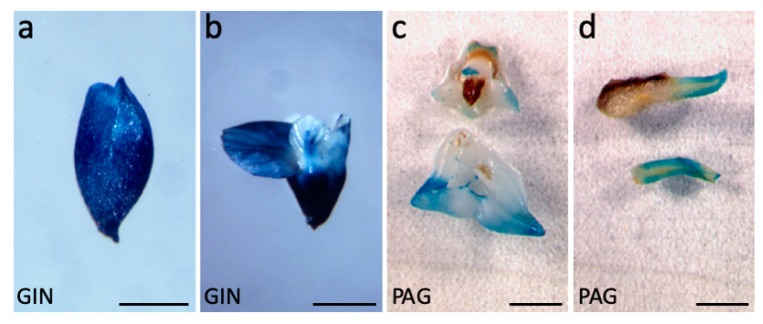
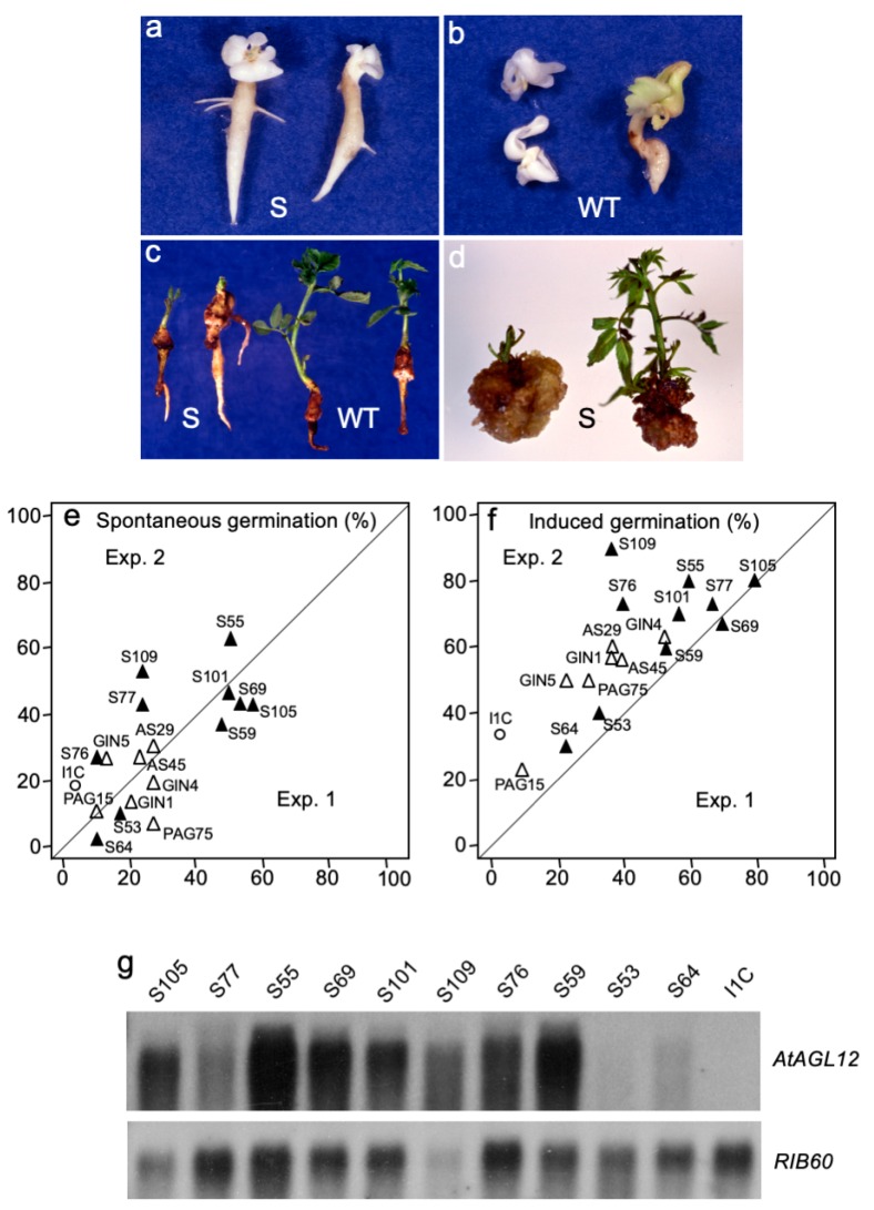
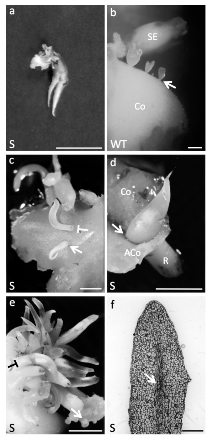
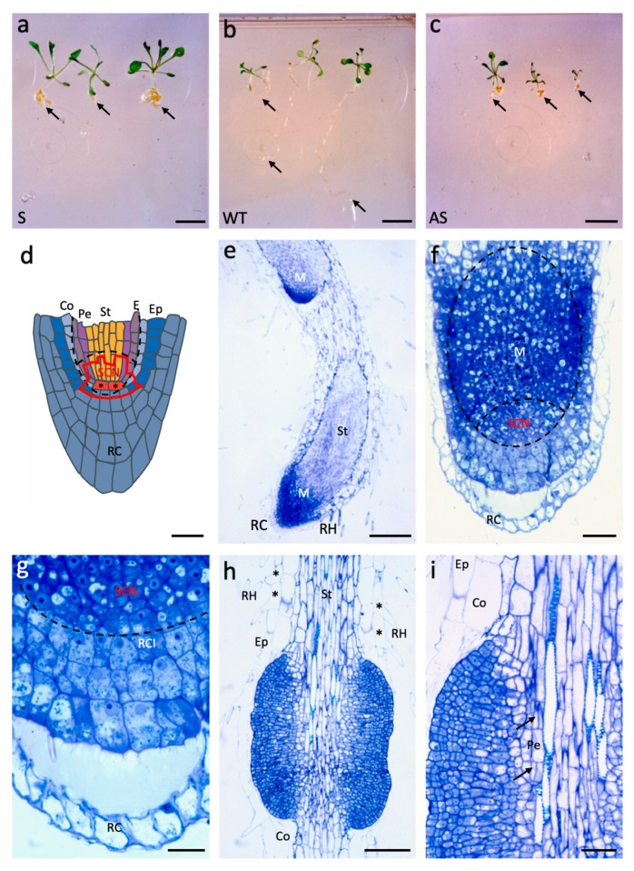
Until recently, the roles of plant MADS-box genes have mainly been characterized during inflorescence and flower differentiation. In order to precise the roles of AGAMOUS-LIKE 12, one of the few MADS-box genes preferentially expressed in roots, we placed its cDNA under the control of the double 35S CaMV promoter to produce transgenic walnut tree and Arabidopsis plants. In Juglans sp., transgenic somatic embryos showed significantly higher germination rates but abnormal development of their shoot apex prevented their conversion into plants. In addition, a wide range of developmental abnormalities corresponding to ectopic root-like structures affected the transgenic lines suggesting partial reorientations of the embryonic program toward root differentiation. In Arabidopsis, AtAGL12 overexpression lead to the production of faster growing plants presenting dramatically wider and shorter root phenotypes linked to increased meristematic cell numbers within the root apex. In the upper part of the roots, abnormal cell divisions patterns within the pericycle layer generated large ectopic cell masses that did not prevent plants to grow. Taken together, our results confirm in both species that AGL12 positively regulates root meristem cell division and promotes overall root vascular tissue formation. Genetic engineering of AGL12 expression levels could be useful to modulate root architecture and development.

Keywords: cell differentiation; cell division; root meristem; transcription factor; transgenic plant.


Conflict of interest statement

The authors declare no conflict of interest.

Figures

Figure 1
Figure 1
Walnut tree somatic embryo transformation. (a) Expression of d35S::GUS in a transgenic early cotyledonary stage walnut somatic embryo (GIN5 line). (b) Expression of d35S::GUS in a mature GIN1 somatic embryo. (c) Patchy expression of pAtAGL12::GUS in the cotyledons of mature somatic embryo (PAG75 line). (d) Expression of pAtAGL12::GUS in the root of a germinated somatic embryo (PAG15 line). Scale bars: 2.5 mm in (a); 5 mm in (bd).
Figure 2
Figure 2
AtAGL12 overexpression in walnut somatic embryos and plantlets. (a) Accelerated spontaneous germination of S101 somatic embryos overexpressing AtAGL12 in sense orientation after 3 weeks of horizontal culture. (b) Wild-type I1C embryos sampled at the same time as control. Note the earlier initiation of lateral root development among the S embryos; (c) conversion of walnut somatic embryos into plantlets. S109 transgenic plantlets (left) and wild type I1C control (right) were photographed after 5 weeks of development in individual tubes. Note the much more important root to shoot ratios for the S plantlets; (d) variable micropropagation and shoot development abilities of the transgenic S lines: the S69 line (left) presenting high AtAGL12 expression levels is characterized by poor shoot apical development and important callus development. Conversely, the S64 line (right) characterized by a low transgene expression levels presents a normal shoot apical development; (e) spontaneous germination rates of representative walnut somatic embryo S, AS, PAG, GIN and wild type I1C lines were scored after 3 weeks of culture. For each line, the numbers of germinated somatic embryos were determined twice (experiment 1 and 2, n = 3 × 10). (f) Induced germination rates were scored after the ablation of the cotyledons and 2 additional weeks of vertical culture (for a total of five weeks of culture). The average germination percentages observed for each S (closed triangles, bold characters), transgenic control (AS, GIN and PAG lines, open triangles) and the wild-type I1C lines (open circle, bold characters) are reported for both experimental repeats and culture times; (g) relative expression levels of AtAGL12 detected in mature non-germinated embryos of the selected walnut transgenic S lines revealed by northern-blot. A walnut Rib60S cDNA probe was used as reference.
Figure 3
Figure 3
Developmental abnormalities linked to AtAGL12 overexpression in walnut. (a) S77 somatic embryo (SE) showing two fused primary roots after 3 weeks of culture. SE presenting fused primary roots were only observed among S embryogenic lines. Scale bar: 1 cm; (b) normal repetitive secondary somatic embryogenesis process in wild-type walnut tree I1C line. The picture shows different stages of SE emerging from the cotyledon (Co) of a primary embryo. The arrow points to the thin basal parts of the secondary embryos that develop from single activated epidermal cells. Scale bar: 1 mm. (c) Ectopic root-like structure (arrow) developing on the apical side of a globular embryo (S77) in place of a regular shoot apex. No cotyledon will form. As in 1b, note the small size of the base of the embryo linked to its single cell origin within the primary cotyledon’s epidermis. When such root-like structures develop directly from activated cells within the epidermal layer, they present a thicker base and need to be cut of the cotyledon in order to be separated from it (T-shaped symbol as in 1e). Scale bar: 0.5 cm; (d) root-like structure developing on the apical part of mature S77 somatic embryo. Excision of the cotyledon reveals the base of the structure originating at the cotyledon/shoot apical meristem junction (arrow). Co: cotyledon, ACo: ablated cotyledon, R: embryonic root pole. Scale bar: 1 cm; (e) cluster of ectopic root-like structures emerging from activated epidermal cells (S76). Clusters of secondary somatic embryos can be observed on the same initial explant (arrow). Scale bar: 0,5 cm; (f) longitudinal section of an ectopic root-like structure (S76). In the central axis of these structures one can observe the initiation of formation of a vascular bundle (arrow) in the absence of a clearly defined apical meristematic zone. Scale bar: 500 μm.
Figure 4
Figure 4
AtAGL12 overexpression in Arabidopsis thaliana. (a) Sense (S) T1 kanR plantlets 10 days after germination; scale bar: 1 cm; (b) wild-type (WT) plantlets 10 days after germination; scale bar: 1 cm (c) Antisense (AS) T1 kanR plantlets 10 days after germination; scale bar: 1 cm; for each plantlet, an arrow points to the apical root tip. (d) Schematic longitudinal section of a WT Arabidopsis root tip adapted from [28]. The main cell types forming the root apical meristem are indicated in different colors: RC: root cap (grey-blue); Ep: epidermis (dark blue); Co: cortex (light blue); E: endodermis (purple-grey), Pe: pericycle (purple); St: Stele (orange). In the stem cell niche (SCN, encircled in red) of the meristem, the quiescent center cells (QC, red with asterisks) are surrounded by a small number of initial cells whose descendants will further divide and differentiate. The main areas of the meristem affected by AtAGL12 overexpression are encircled by dashed lines (see 1f and 1g). Scale bar: 20 μm. (e) Longitudinal section of the primary root tip of a S plantlet presenting two fused roots. In S plantlets, the apical root meristematic zone (M) is enlarged and the stele (St) wider. RH: root hair. Scale bar: 100 μm. (f). Longitudinal section of a S plantlet root tip. The meristematic areas principally affected by AtAGL12 overexpression are encircled (dashed lines). Compared to WT (1d), the basal SNC area of the meristem is wider, characterized by a greater number of meristematic cells. Scale bar: 20 μm. (g) Closer view of the 1f root tip focused on the SNC area. Scale bar: 10 μm; (h) longitudinal section of the upper part of the root of a S plantlet showing numerous cell divisions resulting in the formation of abnormal cell masses around the stele. The presence of adjacent trichoblasts (*) developing root hairs (RH) can be observed. Ep: epidermis, Co: cortex, St: stele. Scale bar: 50 μm. (i) Closer view of the cellular protuberance. The arrows point to closely occurring periclinal cell divisions within the pericycle layer (Pe). Further, both anticlinal and periclinal cell divisions explain the important outgrowth of these structures. Scale bar: 20 μm.

Similar articles

Cited by

References

    1. Yanofsky M.F., Ma H., Bowman J.L., Drews G.N., Feldmann K.A., Meyerowitz E.M. The protein encoded by the Arabidopsis homeotic gene agamous resembles transcription factors. Nature. 1990;346:35–39. doi: 10.1038/346035a0. - DOI - PubMed
    1. Soltis D.E., Soltis P.S., Albert V.A., Oppenheimer D.G., dePamphilis C.W., Ma H., Frohlich M.W., Theiβen G. Missing links: The genetic architecture of flower and floral diversification. Trends Plant Sci. 2002;7:22–31. doi: 10.1016/S1360-1385(01)02098-2. - DOI - PubMed
    1. O’Maoileidigh D.S., Graciet E., Wellmer F. Gene networks controlling Arabidopsis thaliana flower development. New Phytol. 2014;201:16–30. doi: 10.1111/nph.12444. - DOI - PubMed
    1. Alvarez-Buylla E.R., Liljegren S.J., Pelaz S., Gold S.E., Burgeff C., Ditta G.S., Vergara-Silva F., Yanofsky M.F. MADS-box gene evolution beyond flowers: Expression in pollen, endosperm, guard cells, roots and trichomes. Plant J. 2000;24:457–466. doi: 10.1046/j.1365-313x.2000.00891.x. - DOI - PubMed
    1. Leseberg C.H., Li A., Kang H., Duvall M., Mao L. Genome-wide analysis of the MADS-box gene family in Populus trichocarpa. Gene. 2006;378:84–94. doi: 10.1016/j.gene.2006.05.022. - DOI - PubMed

LinkOut - more resources