. 2020 Apr 2;6(2):45.
doi: 10.3390/jof6020045.

Efficacy of Chelerythrine Against Mono- and Dual-Species Biofilms of Candida albicans and Staphylococcus aureus and Its Properties of Inducing Hypha-to-Yeast Transition of C. albicans

Affiliations

Efficacy of Chelerythrine Against Mono- and Dual-Species Biofilms of Candida albicans and Staphylococcus aureus and Its Properties of Inducing Hypha-to-Yeast Transition of C. albicans

Weidong Qian et al. J Fungi (Basel). .

Retraction in

Abstract

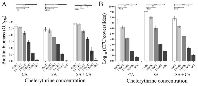
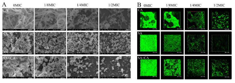
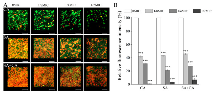
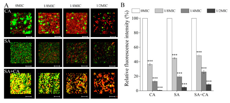
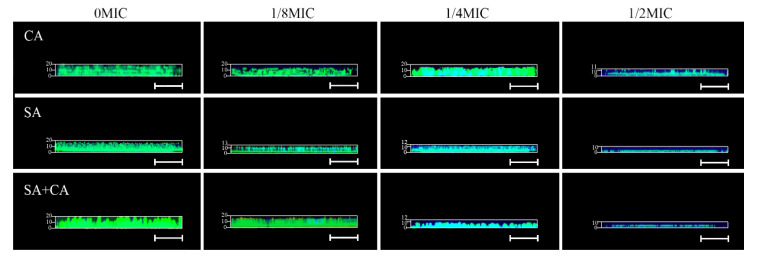
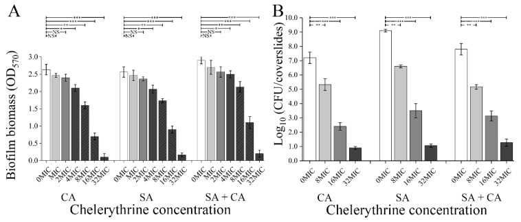
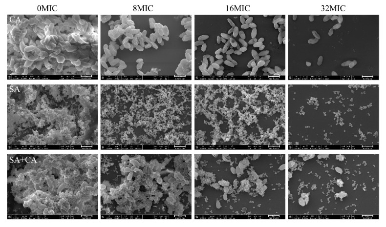
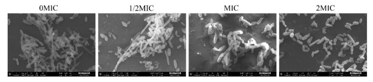
Candida albicans and Staphylococcus aureus specifically often resulted in biofilm-associated diseases, ranging from superficial mucosal to life-threatening systemic infections. Recent studies reported that chelerythrine displayed antimicrobial activities against a few microorganisms, but its effects on mono- and dual-species biofilms of C. albicans and S. aureus have never been reported. The purpose of this study was to evaluate the efficacy of chelerythrine against mono- and dual-species biofilms, and explore its effect on the hyphal growth and the hypha-to-yeast transition of C. albicans. The results showed that minimum inhibitory concentrations (MICs) and minimum biofilm inhibitory concentration (MBIC90S) of chelerythrine against planktonic cells of mono-species were 4 and 2 μg/mL, while the MIC and MBIC90 were 6 and 3 μg/mL for dual-species. Meanwhile, the decrease in three matrix component levels and tolerance to antibiotics of biofilms formed by mono- and dual-species exposed to chelerythrine were confirmed by a confocal laser scanning microscope, in conjugation with five fluorescent dyes and a gatifloxacin diffusion assay. Moreover, C. albicans and S. aureus mono-species showed a 96.4, and 92.3% reduction, respectively, in 24-h preformed biofilm biomass in the presence of 128 µg/mL of chelerythrine. Similarly, preformed (24 h) dual-species biofilm biomass also displayed a significant reduction (90.7%) when treated with 192 μg/mL chelerythrine. Chelerythrine inhibited hyphae formation of C. albicans at 4 μg/mL, and C. albicans in hypha-form can be converted into yeast-form at 8 μg/mL of chelerythrine. Therefore, chelerythrine shows promise as a potential antimicrobial and antibiofilm agent for clinical effective treatments of mono- and mixed-species and/or biofilm-associated infections.

Keywords: Antibiofilm activity; Candida albicans; Chelerythrine; Dual-species biofilm; Staphylococcus aureus.


Conflict of interest statement

The authors declare no conflict of interest.

Figures

Figure 1
Figure 1
Assessment of different concentrations of chelerythrine on C. albicans (CA) and S. aureus (SA) mono- and dual-species (SA+CA) biofilm biomass by crystal violet staining and cell enumeration by colony count. (A) The biofilm biomass was determined using the crystal violet staining assay. Values represent the means of triplicate measurements. (B) Biofilm production analysis was performed by cell enumeration (log10 CFU/coverslips). Bars represent the standard deviation (n = 3). * p < 0.05; ** p < 0.01; *** p < 0.001; NS, not significant.
Figure 2
Figure 2
Inhibitory effects of sub-MIC chelerythrine on C. albicans (CA) and S. aureus (SA) mono- and dual-culture (SA+CA) biofilms, shown in field emission scanning electron microscopy (FESEM, (A)) and confocal laser scanning microscopy images (CLSM, (B)). Scale bars represent 5 μm for FESEM and 10 μm for CLSM, respectively.
Figure 3
Figure 3
Effect of different concentrations of chelerythrine on the eDNA levels within C. albicans (CA) and S. aureus (SA) mono- and dual-species (SA+CA) biofilms by confocal laser scanning microscope. (A) eDNA was labelled with a red fluorescent membrane impermeable DNA-binding stain PI, and bacterial cells were stained with a green-fluorescent nucleic acid counterstain STYO 9. Scale bars represent 10 μm. (B) The relative fluorescence intensity of eDNA in biofilms of each treatment group was plotted against that in untreated group by measuring red fluorescence intensities using KS 400 version 3.0 software. Bars represent the standard deviation (n = 3). *** p < 0.001.
Figure 4
Figure 4
Effect of different concentrations of chelerythrine on C. albicans (CA) and S. aureus (SA) mono- and dual-species (SA+CA) biofilm matrix structure. (A) Extracellular proteins were stained with a red fluorescent stain SYPRO Ruby, and C. albicans and S. aureus cells were stained with a green-fluorescent nucleic acid counterstain STYO 9. Scale bars represent 10 μm. (B) The relative fluorescence intensity of extracellular proteins in biofilms of each treatment group was plotted against that in untreated group by measuring red fluorescence intensities using KS 400 version 3.0 software. Bars represent the standard deviation (n = 3). *** p < 0.001.
Figure 5
Figure 5
Effects of different concentrations of chelerythrine on the levels of extracellular polysaccharides inside C. albicans (CA) and S. aureus (SA) mono- and dual-species (SA+CA) biofilms. (A) Extracellular polysaccharides were stained with a green fluorescent stain wheat germ agglutinin, and C. albicans and S. aureus cells were labelled with a red lipophilic membrane dye FM 4-64. Scale bars represent 10 μm. (B) The relative fluorescence intensity of extracellular polysaccharides in biofilms of each treatment group was plotted against that in untreated group by measuring green fluorescence ratios using KS 400 version 3.0 software. Bars represent the standard deviation (n = 3). *** p < 0.001.
Figure 6
Figure 6
Representative CLSM images assessing diffusion of gatifloxacin within C. albicans (CA) and S. aureus (SA) mono- and dual-species (SA+CA) biofilms formed in the presence of chelerythrine. After mono- and dual-species biofilms were grown for 24 h supplemented without or with chelerythrine of different concentrations, 0.4 mg/mL gatifloxacin was added into the medium. Following 5 h gatifloxacin diffusion, the biofilms were visualized using CLSM. Biofilms were stained with SYTO 9 for biofilms (green) and the intrinsic fluorescence of gatifloxacin (blue). Scale bars represent 10 μm.
Figure 7
Figure 7
Assessment of eradication effects of different concentrations of chelerythrine on C. albicans (CA) and S. aureus (SA) mono- and dual-species (SA+CA) mature biofilm biomass. (A) The biofilm biomass was assessed using crystal violet staining assay. (B) Mature mono and mixed species biofilms were treated with different concentrations of chelerythrine, and fungal or bacterial cell viability was examined by CFU recovery. Values represent the means of triplicate measurements. Bars represent the standard deviation (n = 3). * p < 0.05; ** p < 0.01; *** p < 0.001; NS, not significant.
Figure 8
Figure 8
Scanning electron microscopy images of C. albicans (CA) and S. aureus (SA) mono- and dual-species (SA+CA) mature biofilms after treatment with four MIC-fold concentrations of chelerythrine. Each field of vision was magnified 10,000×. Scale bars represent 5 μm.
Figure 9
Figure 9
The effect of chelerythrine on the hyphal growth of C. albicans. Candida albicans cells were grown in an RPMI-1640 medium at the indicated concentration of chelerythrine at 37 °C for 24 h. Each field of vision was magnified 5000×. Scale bars represent 10 μm.
Figure 10
Figure 10
The effect of chelerythrine on C. albicans mature hyphae. Candida albicans mature hypha were formed in RPMI-1640 for 24 h and treated with indicated concentrations of chelerythrine for 5 h at 37 °C. Each field of vision was magnified 5000×. Scale bars represent 10 μm.

References

    1. Diekema D.J., Messer S.A., Brueggemann A.B., Coffman S.L., Doern G.V., Herwaldt L.A., Pfaller M.A. Epidemiology of candidemia: 3-year results from the emerging infections and the epidemiology of Iowa organisms study. J. Clin. Microbiol. 2002;40:1298–1302. doi: 10.1128/JCM.40.4.1298-1302.2002. - DOI - PMC - PubMed
    1. Magill S.S., O’Leary E., Janelle S.J., Thompson D.L., Dumyati G., Nadle J., Wilson L.E., Kainer M.A., Lynfield R., Greissman S., et al. Changes in prevalence of health care-associated infections in U.S. hospitals. N. Engl. J. Med. 2018;379:1732–1744. doi: 10.1056/NEJMoa1801550. - DOI - PMC - PubMed
    1. Dennis E.K., Garneau-Tsodikova S. Synergistic combinations of azoles and antihistamines against Candida species in vitro. Med. Mycol. 2019;57:874–884. doi: 10.1093/mmy/myy088. - DOI - PubMed
    1. Peters B.M., Jabra-Rizk M.A., O’May G.A., Costerton J.W., Shirtliff M.E. Polymicrobial interactions: Impact on pathogenesis and human disease. Clin. Microbiol. Rev. 2012;25:193–213. doi: 10.1128/CMR.00013-11. - DOI - PMC - PubMed
    1. Pan J., Zhao J., Jiang N. Oral cavity infection: An adverse effect after the treatment of oral cancer in aged individuals. J. Appl. Oral. Sci. 2014;22:261–267. doi: 10.1590/1678-775720130546. - DOI - PMC - PubMed

Publication types

LinkOut - more resources