. 2020 Apr 2;9(4):1009.
doi: 10.3390/jcm9041009.

Immunohistochemical Results of Soft tissues Around a New Implant Healing-Abutment Surface: A Human Study

Affiliations

Immunohistochemical Results of Soft tissues Around a New Implant Healing-Abutment Surface: A Human Study

Barbara Ghinassi et al. J Clin Med. .

Abstract

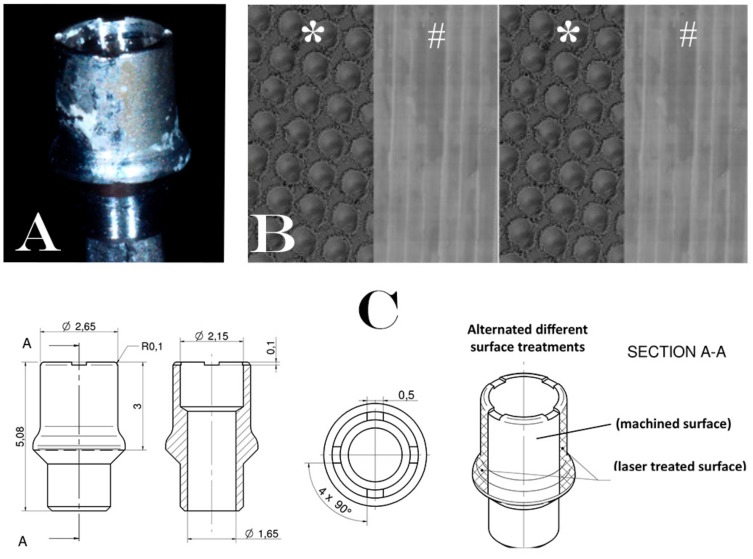
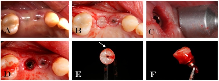
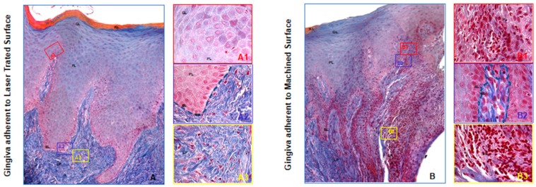
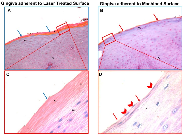
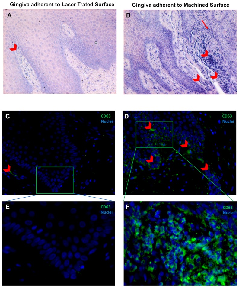
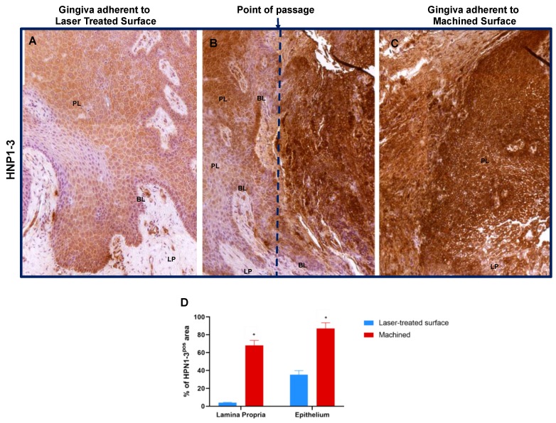
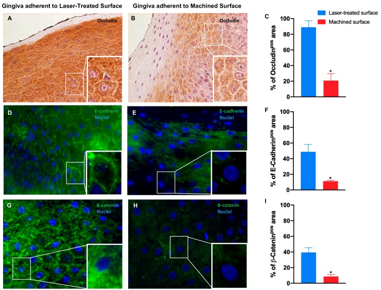
Although, the high success rate of implant rehabilitation treatment, the biological complications such as bone loss and peri-implantitis are still present. The creation of a coronal biological seal between the implant and the oral tissues seems to be a crucial point on preserving dental implants. The objective of this study was to immunohistochemically analyze the behavior of peri-implant soft tissues around a new implant healing-abutment surface on humans. A total of 30 soft tissue biopsies were collected after a healing period of 30 (±7) days, to analyze the expression of inflammatory (cluster of differentiation 63 (CD63), human neutrophil peptides 1-3 (HPN1-3)) and junctional (E-cadherin, occludin, and β-catenin) markers, on soft tissues around laser treated and machined alternated healing abutments. The evaluation demonstrated the whole area of the soft tissues adherent to the laser treated surface with a regular morphology. While several stress hallmarks in correspondence of machined surfaces were shown such as: a) An irregular, disrupted, and discontinued basal membrane with an increased inflammation evident both the epithelial and connective tissues; b) the absence or defective proper keratinization process of the external layer, and c) damages in the cell to cell interaction. In conclusion, the laser treated surface is preferable to maintain the integrity and functionality of the gingiva epithelium.

Keywords: dental implant; immunohistochemical analysis; implant surface; laser treated; peri-implant soft tissues.


Conflict of interest statement

The authors declare that they have no conflict of interest.

Figures

Figure 1
Figure 1
Description of experimental healing abutment used into the study: (A) Healing abutment after the removal of soft tissue. (B) Schematic representation of surfaces treatment on the healing abutment. Each healing abutment was treated with two alternated different surface treatments (laser treated (*)/machined (#)/laser treated (*)/machined (#)); (C) technical design of experimental healing abutment.
Figure 2
Figure 2
Explanatory case of treatment performed: (A) Soft tissue surrounding the experimental healing abutment; (B) circular incision drawing around the healing screw; (C) mucotome used for tissue biopsy; (D) implant after tissue removal; (E) occlusal vision of removal soft tissue and healing abutment. Arrow indicates micro-incision performed in order to know the spatial orientation of biopsy; (F) lateral vision of removal soft tissue and healing abutment.
Figure 3
Figure 3
Trichrome staining of the region of the gingiva adherent to the laser-conditioned- (A) or to machined surfaces (B). In higher magnification of the gingiva adherent to laser treated surface it is evident the regular morphology in all layers: Junctional epithelium (A1), sulcular epithelium with all the typical layers including continuous basal membrane highlighted with the dotted blue line (A2) and subepithelial connective tissue of the lamina propria (A3). In higher magnification of the gingiva adherent to machined surface the typical morphology of the junctional epithelium is not respected (B1): the basal membrane is disrupted (highlighted with the dotted blue line in (B2) and the collagen fibers of subepithelial connective tissue of the lamina propria looks infiltrated with large inflamed area (B3). Magnification 10X (A,B) or 40X (A13,B13). Red, yellow and blue rectangles in A and B represent enlarged areas in the panels on the side, as indicated. Panel A and B are the results of sequential images mounted together. LP: Lamina propria; MB: basilar membrane (dotted blue line); BL: Basal Layer; PL: Prickle layer; GL: granular layer, KL: keratinized layer.
Figure 4
Figure 4
Trichrome staining (A,B) and May–Grünwald–Giemsa (C,D) staining of the region of the gingiva adherent to the laser treated (A,C) or to machined (B,D) surfaces, as indicated. The keratinized layer (that react in orange in the Trichrome staining and pink in the May–Grünwald–Giemsa staining) is barely present or absent (red arrows) in the gingiva adherent to the machined surface and the keratinized layer present still nucleated cells (red arrowheads) or a very thin enucleated cells layer compared to the regular corneal layer of the epithelium of the part of gingiva facing the laser-conditioned surface that present abundant anucleated keratinized layer (blu arrows). Magnification 20X (A,B) or 40X (C,D). Red rectangles in (A,B) represent enlarged areas in (C,D), as indicated PL: prickle layer; GL: granular layer, KL: keratinized layer.
Figure 5
Figure 5
May–Grünwald–Giemsa staining (A,B) and Immunofluorescence against CD63 (green) (C,D) of the soft tissue area adherent to the laser-treated (A,C,E) or to machined (B,D,E) surfaces, as indicated. Red arrowheads indicate flogistic area with leukocites accumulation (A,B), that are also positive to CD63 immunostaining (CF). Nuclei are counterstained with 4’,6-diamino-2-phenylindole DAPI in (C,F). Magnification 10X (A,B), 20X (C,D) or 40X (E,F).
Figure 6
Figure 6
Immunostaining against HNP1–3 of the region of the gingiva adherent to the laser-treated (A), to the point of passage between the two surfaces (B) (dotted blue line represents an arbitrary hypothetical line) and to machined (C) surfaces, as indicated. Magnification 10X. (D) Percentage of HNP1–3 immunostaining positive area in the lamina propria and in the epithelial layers, as indicated. Values are expressed as mean ±SD of five determinations in randomly chosen sections for each specimen. * indicates values statistically different (p < 0.001) between region of the gingiva adherent to the laser-treated- or to machined surfaces, as indicated.
Figure 7
Figure 7
Immunostaining against occludin (A,B) and immunofluorescence for E-cadherin (D,E) and β-catenin (G,H) of the region of gingiva adherent to the laser-treated surface and to machined surface, as indicated. Magnification: 20X (or 40X in the inserts) in (A,B); 40X and (or 100X in the inserts) in (D,E,G). Percentage of occludin (C), E-cadherin (F) and β-catenin (I) positive staining area in the epithelial layers are pointed out in the bar graphs. Values are expressed as mean ±SD of five determinations in randomly chosen sections for each specimen. The (*) indicates values statistically different (p < 0.001) between the area of the soft tissue’s adherent to the laser-treated- or to machined surfaces, as indicated.

References

    1. Simonis P., Dufour T., Tenenbaum H. Long-term implant survival and success: A 10–16-year follow-up of non-submerged dental implants. Clin. Oral Implant. Res. 2010;21:772–777. doi: 10.1111/j.1600-0501.2010.01912.x. - DOI - PubMed
    1. Moraschini V., Poubel L.A., Ferreira V.F., Barboza Edos S. Evaluation of survival and success rates of dental implants reported in longitudinal studies with a follow-up period of at least 10 years: A systematic review. Int. J. Oral Maxillofac. Surg. 2015;44:377–388. doi: 10.1016/j.ijom.2014.10.023. - DOI - PubMed
    1. Misch C.E., Perel M.L., Wang H.L., Sammartino G., Galindo-Moreno P., Trisi P., Steigmann M., Rebaudi A., Palti A., Pikos M.A., et al. Implant success, survival, and failure: The International Congress of Oral Implantologists (ICOI) Pisa Consensus Conference. Implant Dent. 2008;17:5–15. doi: 10.1097/ID.0b013e3181676059. - DOI - PubMed
    1. Berardini M., Trisi P., Sinjari B., Rutjes A.W., Caputi S. The Effects of High Insertion Torque Versus Low Insertion Torque on Marginal Bone Resorption and Implant Failure Rates: A Systematic Review with Meta-Analyses. Implant Dent. 2016;25:532–540. doi: 10.1097/ID.0000000000000422. - DOI - PubMed
    1. Caricasulo R., Malchiodi L., Ghensi P., Fantozzi G., Cucchi A. The influence of implant-abutment connection to peri-implant bone loss: A systematic review and meta-analysis. Clin. Implant Dent. Relat. Res. 2018;20:653–664. doi: 10.1111/cid.12620. - DOI - PubMed

LinkOut - more resources