Review
. 2020 Apr 2;9(4):870.
doi: 10.3390/cells9040870.

Diagnostic and Therapeutic Potential of TSPO Studies Regarding Neurodegenerative Diseases, Psychiatric Disorders, Alcohol Use Disorders, Traumatic Brain Injury, and Stroke: An Update

Affiliations
Review

Diagnostic and Therapeutic Potential of TSPO Studies Regarding Neurodegenerative Diseases, Psychiatric Disorders, Alcohol Use Disorders, Traumatic Brain Injury, and Stroke: An Update

Jasmina Dimitrova-Shumkovska et al. Cells. .

Abstract

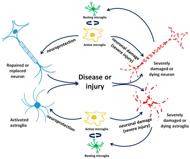
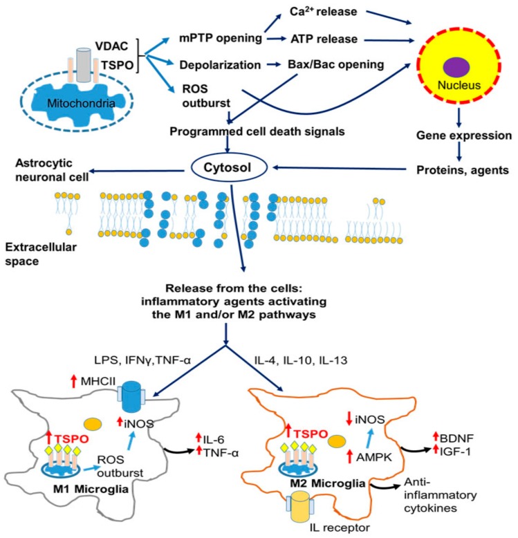
Neuroinflammation and cell death are among the common symptoms of many central nervous system diseases and injuries. Neuroinflammation and programmed cell death of the various cell types in the brain appear to be part of these disorders, and characteristic for each cell type, including neurons and glia cells. Concerning the effects of 18-kDa translocator protein (TSPO) on glial activation, as well as being associated with neuronal cell death, as a response mechanism to oxidative stress, the changes of its expression assayed with the aid of TSPO-specific positron emission tomography (PET) tracers' uptake could also offer evidence for following the pathogenesis of these disorders. This could potentially increase the number of diagnostic tests to accurately establish the stadium and development of the disease in question. Nonetheless, the differences in results regarding TSPO PET signals of first and second generations of tracers measured in patients with neurological disorders versus healthy controls indicate that we still have to understand more regarding TSPO characteristics. Expanding on investigations regarding the neuroprotective and healing effects of TSPO ligands could also contribute to a better understanding of the therapeutic potential of TSPO activity for brain damage due to brain injury and disease. Studies so far have directed attention to the effects on neurons and glia, and processes, such as death, inflammation, and regeneration. It is definitely worthwhile to drive such studies forward. From recent research it also appears that TSPO ligands, such as PK11195, Etifoxine, Emapunil, and 2-Cl-MGV-1, demonstrate the potential of targeting TSPO for treatments of brain diseases and disorders.

Keywords: PET tracing; TSPO; astrocytes; brain disease; brain disorders; cell death; drug development; microglia; microglia activation; neurons; regeneration.


Conflict of interest statement

The authors declare no conflict of interest.

Figures

Figure 1
Figure 1
Principal functions of the 18-kDa translocator protein (TSPO): initiation of programmed cell death, including apoptosis, as well as regulation of the cell’s nuclear gene expression. This figure presents an overview of the cellular pathways influenced by TSPO. TSPO favors neurosteroid synthesis by transporting cholesterol over the outer mitochondrial membrane to the inner mitochondrial membrane. Furthermore, it modulates ATP synthase activity, thus initiating reactive oxygen species (ROS) production, which can result in cardiolipin oxidation and opening of the mitochondrial permeability transition pore (mPTP). Opening of the mPTP allows for ATP and Ca2+ release, and depolarization of the mitochondrial membrane. This depolarization results in opening of the Bax/Bak channel, allowing for the passage of cytochrome c into the cytosol as an initiating step for the mitochondrial apoptosis cascade. The mitochondrial ROS generation and ATP and Ca2+ release are also considered part of the mitochondria-to-nucleus pathway of cell nuclear gene expression modulation, which includes the modulation of hundreds to thousands of genes (see also [8,11,12,13]).
Figure 2
Figure 2
Interactions between brain cell types taking part in acute and progressing brain damage after brain injury and disease. Acute microglial activation following mild brain damage causes may provide protection to neurons and astrocytes while long-term glial activation may cause damage to and the death of neurons and astrocytes.
Figure 3
Figure 3
Cellular pathology of neurodegenerative and neurodevelopmental diseases and its correlation with microglial response and modulation of TSPO ligand-binding potentials. Neurodegeneration initiated by cellular protein modification or aggregation induces an inflammatory response, accompanied by activation of the M1 and/or M2 microglial pathway. Proinflammatory (M1) microglial phenotype activated by LPS, Tumor necrosis factor alpha TNF-α, or Interferon gamma IFNγ stimulation is characterized by the expression of inducible nitric oxide synthase iNOS, MHC II, and significantly increased TSPO BP as well as ROS overproduction and secretion of injury-mediated cytokines. The anti-inflammatory (M2) microglial phenotype is stimulated by interleukins (IL-4, IL-10, IL-13) characterized by the secretion of brain-derived neurotrophic factor (BDNF), Insulin growth factor 1 (IGF-1), and anti-inflammatory cytokines [52]. Abbreviations: AD—Alzheimer disease; AMPK—5′ AMP-activated protein kinase; BDNF—brain derived neutrophic factor; BPAD—Bipolar affective disorder; HD—Huntington disease; IFNγ—Interferon gamma; IGF1—Insulin growth factor 1; IL—Interleukin; iNOS—Inducible nitric oxide synthase; LPS—Lipopolysaccharide; MDD—Major depressive disorder; MHCII—Major histocompatibility complex protein class II; PD—Parkinson disease; SCZ—Schizophrenia; TNF-α—Tumor necrosis factor alpha.
Figure 4
Figure 4
Intracellular manifestation of TBI in neurons and astrocytes, resulting in microglial activation. A vicious cycle of cell death and activation associated with enhanced TSPO responses’ expression in brain injury is reflected by cellular apoptosis and necrosis attained by way of mPTP opening and a decrease of intraneuronal ATP, which consequently triggers the neuroinflammatory response by microglial activation. Proinflammatory (M1) microglial phenotype activated by LPS, TNF-α, or IFNγ stimulation, and characterized by the expression of iNOS, MHCII, and significantly increased TSPO BP, contribute to ROS overproduction and the secretion of injury-mediated cytokines. The anti-inflammatory (M2) microglial phenotype is stimulated by interleukins (IL-4, IL-10, IL-13) and characterized by the secretion of BDNF, IGF-1, and anti-inflammatory cytokines. On the one hand, based on our various studies, we arrive a the assumption that mitochondrial membrane depolarization leads to programmed cell death; on the other hand, less pronounced effects on the mitochondrial membrane potential, but still allowing for ROS generation, and Ca2+ and ATP release into the cytosol, will lead to gene expression modulation that is part of: Microglial activation, astrocytic activation, neuronal and astrocytic cell death, neuronal (re) generation, angiogenesis, and wound healing [13]. Abbreviations: AD—Alzheimer’s disease; AMPK-5′—AMP-activated protein kinase; BDNF—brain derived neutrophic factor; BPAD—Bipolar affective disorder; HD—Huntington’s disease; IFNγ—Interferon gamma; IGF1—Insulin growth factor 1; IL—Interleukin; iNOS—Inducible nitric oxide synthase; LPS—Lipopolysaccharide; MDD—Major depressive disorder; MHCII—Major histocompatibility complex protein class II; mPTP—Mitochondrial permeability transition pore; PD—Parkinson’s disease; SCZ—Schizophrenia; TNF-α—Tumor necrosis factor alpha.

Similar articles

Cited by

References

    1. Gavish M., Bachman I., Shoukrun R., Katz Y., Veenman L., Weisinger G., Weizman A. Enigma of the peripheral benzodiazepine receptor. Pharmacol. Rev. 1999;51:629–650. - PubMed
    1. Papadopoulos V., Lecanu L., Brown R.C., Han Z., Yao Z.X. Peripheral-type benzodiazepine receptor in neurosteroid biosynthesis, neuropathology and neurological disorders. Neuroscience. 2006;138:749–756. doi: 10.1016/j.neuroscience.2005.05.063. - DOI - PubMed
    1. Gavish M., Weizman R. Role of peripheral-type benzodiazepine receptors in steroidogenesis. Clin. Neuropharmacol. 1997;20:473–481. doi: 10.1097/00002826-199712000-00001. - DOI - PubMed
    1. Murphy M.P. How mitochondria produce reactive oxygen species. Biochem. J. 2009;417:1–13. doi: 10.1042/BJ20081386. - DOI - PMC - PubMed
    1. Dimitrova-Shumkovska J., Veenman L., Ristoski T., Leschiner S., Gavish M. Dimethylbenz[alpha]anthracene induces oxidative stress and reduces the binding capacity of the mitochondrial 18-kDa translocator protein in rat aorta. Drug Chem. Toxicol. 2010;33:337–347. doi: 10.3109/01480540903483441. - DOI - PubMed