. 2020 Apr 2;21(7):2476.
doi: 10.3390/ijms21072476.

Impact of Type 2 Diabetes Mellitus on Human Bone Marrow Stromal Cell Number and Phenotypic Characteristics

Affiliations

Impact of Type 2 Diabetes Mellitus on Human Bone Marrow Stromal Cell Number and Phenotypic Characteristics

Féaron C Cassidy et al. Int J Mol Sci. .

Abstract

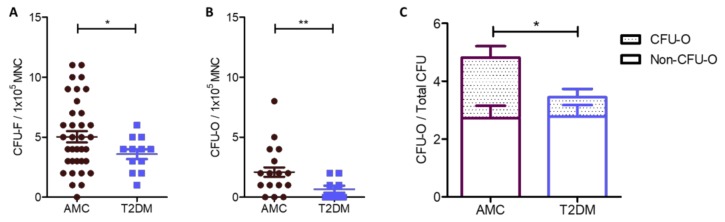
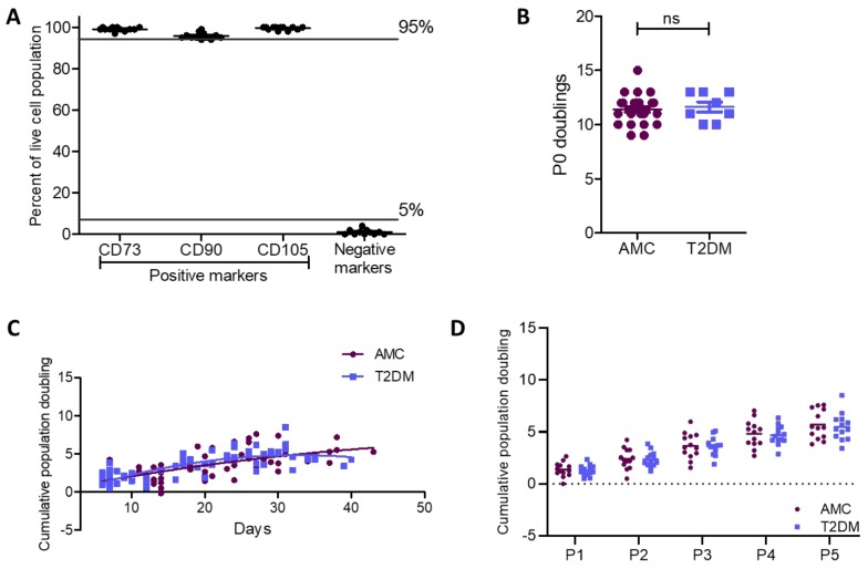
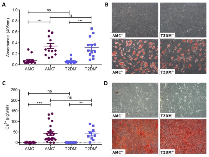
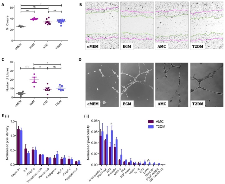
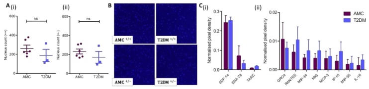
Human bone marrow-derived mesenchymal stromal cells (MSCs) have been investigated in numerous disease settings involving impaired regeneration because of the crucial role they play in tissue maintenance and repair. Considering the number of comorbidities associated with type 2 diabetes mellitus (T2DM), the hypothesis that MSCs mediate these comorbidities via a reduction in their native maintenance and repair activities is an intriguing line of inquiry. Here, it is demonstrated that the number of bone marrow-derived MSCs in people with T2DM was reduced compared to that of age-matched control (AMC) donors and that this was due to a specific decrease in the number of MSCs with osteogenic capacity. There were no differences in MSC cell surface phenotype or in MSC expansion, differentiation, or angiogenic or migratory capacity from donors living with T2DM as compared to AMCs. These findings elucidate the basic biology of MSCs and their potential as mediators of diabetic comorbidities, especially osteopathies, and provide insight into donor choice for MSC-based clinical trials. This study suggests that any role of bone marrow MSCs as a mediator of T2DM comorbidity is likely due to a reduction in the osteoprogenitor population size and not due to a permanent alteration to the MSCs' capacity to maintain tissue homeostasis through expansion and differentiation.

Keywords: adult stem cells; bone marrow stromal cells; mesenchymal stem cells; mesenchymal stromal cells; type 2 diabetes mellitus.


Conflict of interest statement

The authors declare no conflict of interest to disclose with the exception of Prof. Timothy O’Brien who is a founder, director, and equity holder in Orbsen Therapeutics. The funders had no role in the design of the study; in the collection, analyses, or interpretation of data; in the writing of the manuscript; or in the decision to publish the results.

Figures

Figure 1
Figure 1
Quantification of the number of MSCs, detected as CFU-Fs, and those MSCs with native osteogenic potential (CFU-O) within whole marrow aspirates from AMC and T2DM cohorts. (A) Bone marrow from donors living with T2DM (n = 39) contained reduced numbers of CFU-Fs compared to bone marrow from the AMC cohort (n = 12). These data were normalised to MNC number and indicate the number of MSCs in the bone marrow is reduced in response to the T2DM environment. (B) CFU-O numbers in fresh bone marrow were identified by alkaline phosphatase staining and normalised to MNC number as per CFU-F. A reduction in the number of CFU-Os was recorded in the bone marrow in the T2DM cohort (n = 9) compared to that from the AMC cohort (n = 22). (C) In evaluating CFU-O number as a proportion of all CFUs in a donor bone marrow by two-way factorial analysis of deviance, diabetic status was observed to lead to a reduction in the proportion of CFU-Os. All data are displayed as mean ± SEM. * = p ≤ 0.05; ** = p ≤ 0.01.
Figure 2
Figure 2
Cell surface phenotype and proliferative capacity were retained in MSCs from donors with T2DM as compared to AMCs. (A) Flow cytometry confirmed the population of bone marrow-derived primary cells isolated by plastic adherence as being MSCs due to their positive surface marker expression profile (CD73+, CD90+, and CD105+) and absence of CD34, CD45, CD11b or CD14, CD19 or CD79α, and HLA-DR (negative cocktail) expression (n = 12). (B) Population doubling at P0 was unaffected by the diabetic status of the donor (n = 28 for AMC and n = 8 for T2DM cohorts). (C) There was also no difference in doubling capacity between MSCs derived from individuals with T2DM or AMCs when cumulative population doublings per day were compared between the two groups (comparison of second-order polynomial nonlinear regression of the proliferation curve for each cohort of n = 13). (D) A 2-way ANOVA determined that there was no effect of T2DM on proliferative capacity per passage (P1–P5). All data are displayed as mean ± SEM. ns = not significant, i.e., p > 0.05.
Figure 3
Figure 3
Adipogenic and osteogenic differentiation of MSC cultures. Undifferentiated cultures are identified as AMC or T2DM, while differentiated cultures are identified as AMC+ or T2DM+. (A) MSC cultures were induced to form adipocytes, while undifferentiated cultures served as a control. Quantification of Oil Red O stain retention indicates a near absence of absorbed stain in undifferentiated cultures; then a statistically significantly enhanced retention in differentiated cultures. The presence of T2DM in the donor did not influence the culture’s capacity to undergo adipogenic differentiation (n = 13 AMC and n = 14 T2DM donor samples). (B) Representative images of each donor cohort demonstrating Oil Red O positive cells in differentiated cultures at the same frequency in cultures isolated from individuals with T2DM as in AMC cultures. Images obtained by 4× objective as described in methods. (C) Quantification of extracted calcium from osteogenically differentiated MSC cultures indicated their bone-forming potential. Calcium was present at negligible levels in undifferentiated AMC and T2DM control cultures but was significantly enhanced in differentiated cultures. There was no statistically significant impact of T2DM on the capacity of these cells to osteogenically differentiate (n = 23 for AMC and n = 9 for T2DM cohorts). (D) Staining of osteogenically differentiated cultures with Alizarin Red revealed the presence of calcium in the culture’s extracellular matrix. Undifferentiated controls were negative for Alizarin Red staining, while differentiated cultures retained stain. The distribution and intensity of the stain was comparable between donor cohorts. Images obtained by 4× objective as described in methods. All data are displayed as mean ± SEM. * = p ≤ 0.05; ** = p ≤ 0.01, ns = not significant, i.e., p > 0.05.
Figure 4
Figure 4
The presence of T2DM does not influence the capacity of MSC-secreted factors to support angiogenesis. The angiogenic capacity of MSCs to support migration of surrounding cells and to induce tubule formation was evaluated. (A) Quantification of HUVEC cell migration into a scratch in the HUVEC monolayer in the presence of MSC-conditioned medium indicated cells isolated from donors with (n = 10) or without T2DM (n = 8) were capable of stimulating HUVECs to close an in vitro wound. (B) Representative images display a notable decrease in the scratch width as a result of exposure to MSC-conditioned medium regardless of donor cohort. Images obtained by 10× objective as described in methods. Zero h scratch width is overlain onto the photograph taken at 8.5 h, with the 0 h outline traced in purple and the 8.5 h line outlined in green. (C) MSC-conditioned medium was capable of stimulating the formation of endothelial cell tubules regardless of its donor origin (AMC n = 7 and T2DM n = 5). (D) Representative images indicate the presence of long, branching tubules forming complete loops with a comparable morphology in cultures exposed to both types of MSC conditioned medium. Images obtained by 10× objective as described in methods. (E) Angiogenic protein levels in conditioned media at higher concentrations (i) and at lower concentrations (ii). Multiple T-test in R demonstrated increased levels of two secreted proteins (HGF and DPP4, mean ± SEM were 0.0322 ± 0.008 (AMC) and 0.0619 ± 0.009 (T2DM), and −0.002 ± 0.001 (AMC) and 0.003 ± 0.001 (T2DM), respectively), and no significant difference in levels where no statistics summary label is provided on the image (n = 4 per cohort). All data are displayed as mean ± SEM. * = p ≤ 0.05; ** = p ≤ 0.01, *** = p ≤ 0.001, ns = not significant, i.e., p > 0.05.
Figure 5
Figure 5
MSC mobility is not impacted by the diabetic status of the marrow donor. (A) As assayed through a transwell culture system, the (i) chemokinetic and (ii) chemotactic migration of MSCs from AMC (n = 6) and T2DM (n = 3) cohorts are comparable. (B) Representative images demonstrate the underside of a transwell with the migrated nuclei stained with Hoechst. (C) Chemokine levels in conditioned media at higher concentrations (i) and at lower concentrations (ii). Multiple T-test in R demonstrated no significant difference in levels (n = 4 per cohort). All data are displayed as mean + SEM, ns = not significant, i.e., p > 0.05.

References

    1. Dominici M., Le Blanc K., Mueller I., Slaper-Cortenbach I., Marini F., Krause D.S., Deans R.J., Keating A., Prockop D.J., Horwitz E.M. Minimal criteria for defining multipotent mesenchymal stromal cells. The International Society for Cellular Therapy position statement. Cytotherapy. 2006;8:315–317. doi: 10.1080/14653240600855905. - DOI - PubMed
    1. Consentius C., Mirenska A., Jurisch A., Reinke S., Scharm M., Zenclussen A.C., Hennig C., Volk H.-D. In situ detection of CD73+ CD90+ CD105+ lineage: Mesenchymal stromal cells in human placenta and bone marrow specimens by chipcytometry. Cytom. Part A. 2018;93:889–893. doi: 10.1002/cyto.a.23509. - DOI - PubMed
    1. Zhu Y., Feng X. Genetic contribution to mesenchymal stem cell dysfunction in systemic lupus erythematosus. Stem Cell Res. Ther. 2018;9:149. doi: 10.1186/s13287-018-0898-x. - DOI - PMC - PubMed
    1. Rennert R.C., Sorkin M., Januszyk M., Duscher D., Kosaraju R., Chung M.T., Lennon J., Radiya-Dixit A., Raghvendra S., Maan Z.N., et al. Diabetes impairs the angiogenic potential of adipose-derived stem cells by selectively depleting cellular subpopulations. Stem Cell Res. Ther. 2014;5:79. doi: 10.1186/scrt468. - DOI - PMC - PubMed
    1. Kim H., Han J.W., Lee J.Y., Choi Y.J., Sohn Y.-D., Song M., Yoon Y.-S. Diabetic Mesenchymal Stem Cells Are Ineffective for Improving Limb Ischemia Due to Their Impaired Angiogenic Capability. Cell Transplant. 2015;24:1571–1584. doi: 10.3727/096368914X682792. - DOI - PMC - PubMed