

Convolutional Neural Network Technology in Endoscopic Imaging: Artificial Intelligence for Endoscopy
- PMID: 32252504
- PMCID: PMC7137563
- DOI: 10.5946/ce.2020.054
Convolutional Neural Network Technology in Endoscopic Imaging: Artificial Intelligence for Endoscopy
Abstract
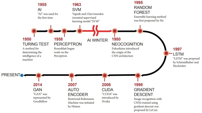
Recently, significant improvements have been made in artificial intelligence. The artificial neural network was introduced in the 1950s. However, because of the low computing power and insufficient datasets available at that time, artificial neural networks suffered from overfitting and vanishing gradient problems for training deep networks. This concept has become more promising owing to the enhanced big data processing capability, improvement in computing power with parallel processing units, and new algorithms for deep neural networks, which are becoming increasingly successful and attracting interest in many domains, including computer vision, speech recognition, and natural language processing. Recent studies in this technology augur well for medical and healthcare applications, especially in endoscopic imaging. This paper provides perspectives on the history, development, applications, and challenges of deep-learning technology.
Keywords: Artificial intelligence; Convolutional neural network; Deep learning; Endoscopic imaging; Machine learning.
Conflict of interest statement
Figures

Similar articles
-
Deep Learning in Medical Imaging: General Overview.Korean J Radiol. 2017 Jul-Aug;18(4):570-584. doi: 10.3348/kjr.2017.18.4.570. Epub 2017 May 19. Korean J Radiol. 2017. PMID: 28670152 Free PMC article. Review.
-
Deep Learning in Medical Imaging.Neurospine. 2019 Dec;16(4):657-668. doi: 10.14245/ns.1938396.198. Epub 2019 Dec 31. Neurospine. 2019. PMID: 31905454 Free PMC article.
-
Survey on Deep Neural Networks in Speech and Vision Systems.Neurocomputing (Amst). 2020 Dec 5;417:302-321. doi: 10.1016/j.neucom.2020.07.053. Epub 2020 Jul 26. Neurocomputing (Amst). 2020. PMID: 33100581 Free PMC article.
-
Deep convolutional neural network and IoT technology for healthcare.Digit Health. 2024 Jan 17;10:20552076231220123. doi: 10.1177/20552076231220123. eCollection 2024 Jan-Dec. Digit Health. 2024. PMID: 38250147 Free PMC article.
-
Deep Learning: A Primer for Radiologists.Radiographics. 2017 Nov-Dec;37(7):2113-2131. doi: 10.1148/rg.2017170077. Radiographics. 2017. PMID: 29131760 Review.
Cited by
-
Feasibility of Deep Learning Algorithms for Reporting in Routine Spine Magnetic Resonance Imaging.Int J Spine Surg. 2020 Dec;14(s3):S86-S97. doi: 10.14444/7131. Int J Spine Surg. 2020. PMID: 33298549 Free PMC article.
-
Usefulness of an artificial intelligence-based colonoscopy report generation support system.Clin Endosc. 2025 Mar;58(2):327-330. doi: 10.5946/ce.2024.213. Epub 2025 Feb 19. Clin Endosc. 2025. PMID: 40010702 Free PMC article. No abstract available.
-
A New Active Locomotion Capsule Endoscopy under Magnetic Control and Automated Reading Program.Clin Endosc. 2020 Jul;53(4):395-401. doi: 10.5946/ce.2020.127. Epub 2020 Jul 30. Clin Endosc. 2020. PMID: 32746536 Free PMC article.
-
Colonoscopic image synthesis with generative adversarial network for enhanced detection of sessile serrated lesions using convolutional neural network.Sci Rep. 2022 Jan 7;12(1):261. doi: 10.1038/s41598-021-04247-y. Sci Rep. 2022. PMID: 34997124 Free PMC article.
-
Future of Endoscopy in Inflammatory Bowel Diseases (IBDs).Cureus. 2022 Sep 25;14(9):e29567. doi: 10.7759/cureus.29567. eCollection 2022 Sep. Cureus. 2022. PMID: 36312686 Free PMC article. Review.
References
-
- McCarthy J, Minsky ML, Rochester N, Shannon CE. A proposal for the dartmouth summer research project on artificial intelligence, August 31, 1955. AI Mag. 2006;27:12–14.
-
- Cortes C, Vapnik V. Support-vector networks. Mach Learn. 1995;20:273–297.
-
- Freund Y, Schapire RE. A decision-theoretic generalization of on-line learning and an application to boosting. J Comput Syst Sci. 1997;55:119–139.
-
- Perronnin F, Sánchez J, Mensink T. Improving the Fisher kernel for large-scale image classification. In: 11th European Conference on Computer Vision; 2010 Sep 5-11; Heraklion, Greece. Berlin. 2010. pp. 143–156.
-
- Krizhevsky A, Sutskever I, Hinton GE. ImageNet classification with deep convolutional neural networks. Advances in Neural Information Processing Systems 25 (NIPS 2012) 2012:1097-1105.
LinkOut - more resources
Full Text Sources
