. 2020 May 1;177(5):454-463.
doi: 10.1176/appi.ajp.2019.19060650. Epub 2020 Apr 7.

Age Differences in the Neural Correlates of Anxiety Disorders: An fMRI Study of Response to Learned Threat

Affiliations

Age Differences in the Neural Correlates of Anxiety Disorders: An fMRI Study of Response to Learned Threat

Andrea L Gold et al. Am J Psychiatry. .

Abstract

Objective: Although both pediatric and adult patients with anxiety disorders exhibit similar neural responding to threats, age-related differences have been found in some functional MRI (fMRI) studies. To reconcile disparate findings, the authors compared brain function in youths and adults with and without anxiety disorders while rating fear and memory of ambiguous threats.

Methods: Two hundred medication-free individuals ages 8-50 were assessed, including 93 participants with an anxiety disorder. Participants underwent discriminative threat conditioning and extinction in the clinic. Approximately 3 weeks later, they completed an fMRI paradigm involving extinction recall, in which they rated their levels of fear evoked by, and their explicit memory for, morph stimuli with varying degrees of similarity to the extinguished threat cues.

Results: Age moderated two sets of anxiety disorder findings. First, as age increased, healthy subjects compared with participants with anxiety disorders exhibited greater amygdala-ventromedial prefrontal cortex (vmPFC) connectivity when processing threat-related cues. Second, age moderated diagnostic differences in activation in ways that varied with attention and brain regions. When rating fear, activation in the vmPFC differed between the anxiety and healthy groups at relatively older ages. In contrast, when rating memory for task stimuli, activation in the inferior temporal cortex differed between the anxiety and healthy groups at relatively younger ages.

Conclusions: In contrast to previous studies that demonstrated age-related similarities in the biological correlates of anxiety disorders, this study identified age differences. These findings may reflect this study's focus on relatively late-maturing psychological processes, particularly the appraisal and explicit memory of ambiguous threat, and inform neurodevelopmental perspectives on anxiety.

Keywords: Age; Anxiety Disorders; Neural Correlates; fMRI.


Figures

FIGURE 1.
FIGURE 1.. Task paradigm of threat conditioning, extinction, and extinction recall among youths and adults with anxiety disorders and healthy subjectsa
a In visit 1 (panel A), participants first underwent threat conditioning, during which one female face (conditioned stimulus [CS+]) was paired with a fearful face coterminating with a loud scream (unconditioned stimulus); the other female face (CS−) was never paired with the unconditioned stimulus. Next, during extinction, the two faces were repeatedly presented without the unconditioned stimulus. In visit 2 (panel B), participants completed extinction recall, during which morphed images continuously varying in similarity from the CS− to CS+ (bottom) were presented. For each image, participants rated (top) their current levels of fear (threat appraisal) or whether the CS screamed in the past (explicit memory).
FIGURE 2.
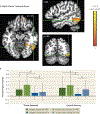
FIGURE 2.. Task-related activation in the ventromedial prefrontal cortex among youths and adults with anxiety disorders and healthy subjectsa
a Whole-brain analyses of task-related activation revealed a significant interaction of anxiety diagnosis, age, and attention condition in the ventromedial prefrontal cortex (vmPFC) (panelA). Images are shown in neurological convention (i.e., left is left) and thresholded at F>10.76, df=1, 4180, p<0.001, cluster size >57 voxels (890.625 mm3). To decompose the complex interaction effects, mean extracted values (panel B) for this cluster are plotted separately by attention condition (threat appraisal, explicit memory) and group, based on anxiety diagnosis (healthy, anxiety) and age (median split: adults, youths). In the graph, the y-axis shows extracted vmPFC percent signal change averaged across participants in each group. Error bars indicate standard deviation. *p<0.05.
FIGURE 3.
FIGURE 3.. Task-related activation in the right inferior temporal gyrus among youths and adults with anxiety disorders and healthy subjectsa
a Whole-brain analyses of task-related activation revealed a significant interaction of anxiety diagnosis, age, and attention condition in two clusters in the right inferior temporal gyrus (ITG) (panel A). Images are shown in neurological convention (i.e., left is left) and thresholded at F>10.76, df=1, 4180, p<0.001, cluster size >57 voxels (890.625 mm3). To decompose the complex interaction effects, mean extracted values (panel B) from the larger ITG cluster (263 voxels) are plotted separately by attention condition (threat appraisal, explicit memory) and group, based on anxiety diagnosis (healthy, anxiety) and age (median split: adults, youths). In the graph, the y-axis shows extracted ITG percent signal change averaged across participants in each group. Error bars indicate standard deviation. *p<0.05.
FIGURE 4.
FIGURE 4.. Left amygdala connectivity with the ventromedial prefrontal cortex (vmPFC) among youths and adults with anxiety disorders and healthy subjectsa
a Whole-brain generalized psychophysiological interaction (gPPI) analysis revealed a significant interaction of anxiety diagnosis, age, and linear trend in the task-related functional connectivity between the left amygdala seed and the vmPFC (panel A). Images are shown in neurological convention (i.e., left is left) and thresholded at F>10.76, df=1, 4180, p<0.001, cluster size >57 voxels (890.625 mm3). To decompose the complex interaction effects, mean extracted values for the vmPFC cluster are plotted separately by group, based on anxiety diagnosis (healthy, anxiety) and age (median split: adults, youths). In the graph (panel B), the y-axis shows the linear trend of the gPPI coefficient averaged across participants in each group. Error bars indicate standard deviation. *p<0.05.

Comment in

Similar articles

Cited by

References

    1. Hardee JE, Benson BE, Bar-Haim Y, et al.: Patterns of neural connectivity during an attention bias task moderate associations between early childhood temperament and internalizing symptoms in young adulthood. Biol Psychiatry 2013; 74:273–279 - PMC - PubMed
    1. White LK, Sequeira S, Britton JC, et al.: Complementary features of attention bias modification therapy and cognitive-behavioral therapy in pediatric anxiety disorders. Am J Psychiatry 2017; 174: 775–784 - PMC - PubMed
    1. Beesdo K, Knappe S, Pine DS: Anxiety and anxiety disorders in children and adolescents: developmental issues and implications for DSM-V. Psychiatr Clin North Am 2009; 32:483–524 - PMC - PubMed
    1. Britton JC, Grillon C, Lissek S, et al.: Response to learned threat: an FMRI study in adolescent and adult anxiety. Am J Psychiatry 2013; 170:1195–1204 - PMC - PubMed
    1. Gold AL, Shechner T, Farber MJ, et al.: Amygdala-cortical connectivity: associations with anxiety, development, and threat. Depress Anxiety 2016; 33:917–926 - PMC - PubMed

Publication types