

Uncrossed corticospinal tracts in a patient with ichthyosis and hemiparesis: a case report
- PMID: 32252685
- PMCID: PMC7132884
- DOI: 10.1186/s12883-020-01698-0
Uncrossed corticospinal tracts in a patient with ichthyosis and hemiparesis: a case report
Abstract
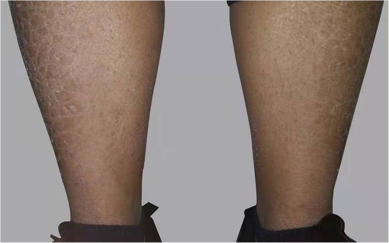
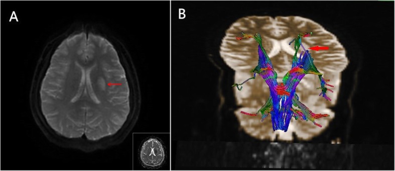
Background: Anomalies of pyramidal tract decussation are rare phenomena that can be caused by ectodermal dysplasia. Herein, we describe a patient with ichthyosis who exhibited ipsilateral hemiparesis after stroke and whose neuroimaging results showed evidence of motor control being provided by the ipsilateral motor cortex.
Case presentation: A 24-year-old right-handed man presented with skin abnormalities, sudden-onset left hemiparesis, and dysarthria. He exhibited a mild-to-moderate left-sided weakness (grade 4 on the Medical Research Council scale). Magnetic resonance imaging revealed an acute infarct in the left corona radiata. Diffusion tensor imaging revealed uncrossed corticospinal tracts. Next-generation sequencing identified heterozygous FLG mutations. The patient was diagnosed with cerebral infarction and ichthyosis vulgaris and was treated with aspirin (100 mg/d). His symptoms gradually dissipated.
Conclusions: This case suggests that pyramidal decussation anomalies can be associated with ichthyosis. Patients with ichthyosis should therefore be evaluated for nerve involvement.
Keywords: DTI; Ihthyosis; Ipsilateral hemiparesis; Ischemic stroke; Uncrossed corticospinal tracts.
Conflict of interest statement
The authors declare that they have no competing interests.
Figures
References
MeSH terms
Substances
Grants and funding
LinkOut - more resources
Full Text Sources
Medical
Miscellaneous
