

CT imaging changes of corona virus disease 2019(COVID-19): a multi-center study in Southwest China
- PMID: 32252784
- PMCID: PMC7132551
- DOI: 10.1186/s12967-020-02324-w
CT imaging changes of corona virus disease 2019(COVID-19): a multi-center study in Southwest China
Abstract
Background: Since the first case of a coronavirus disease 2019 (COVID-19) infection pneumonia was detected in Wuhan, China, a series of confirmed cases of the COVID-19 were found in Southwest China. The aim of this study was to describe the imaging manifestations of hospitalized patients with confirmed COVID-19 infection in southwest China.
Methods: In this retrospective study, data were collected from 131 patients with confirmed coronavirus disease 2019 (COVID-19) from 3 Chinese hospitals. Their common clinical manifestations, as well as characteristics and evolvement features of chest CT images, were analyzed.
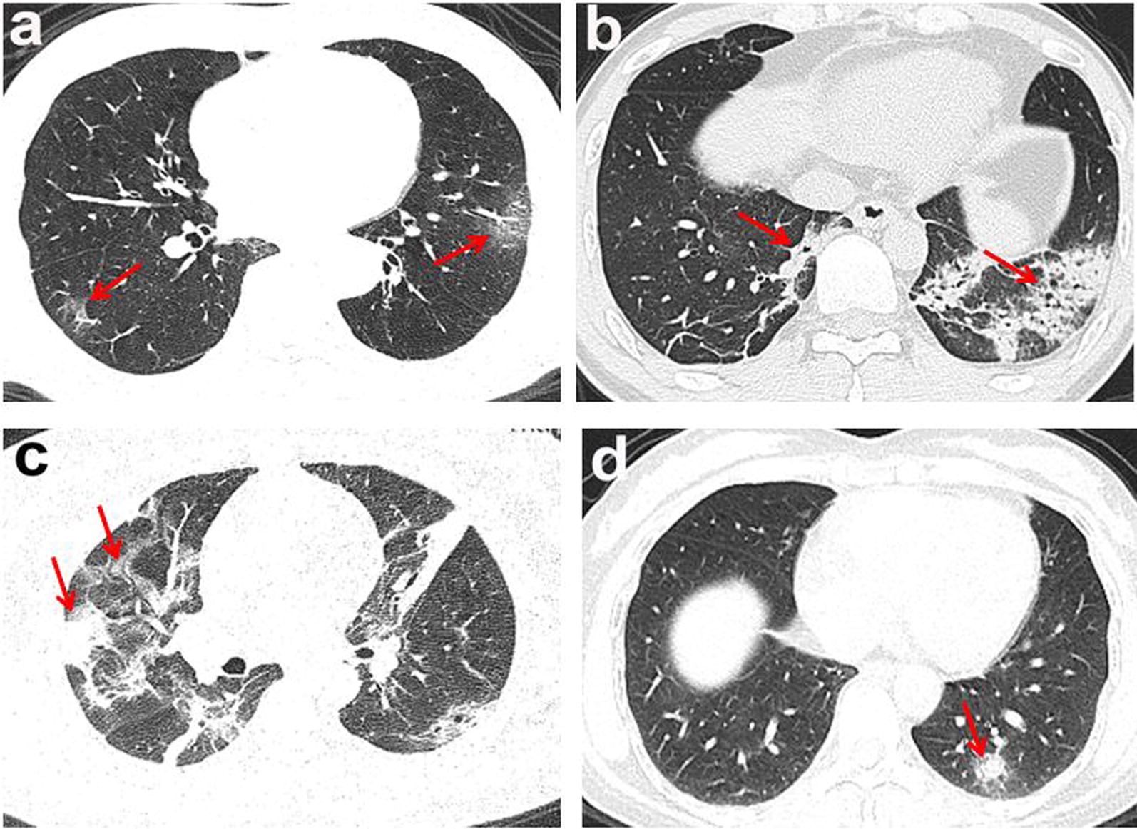
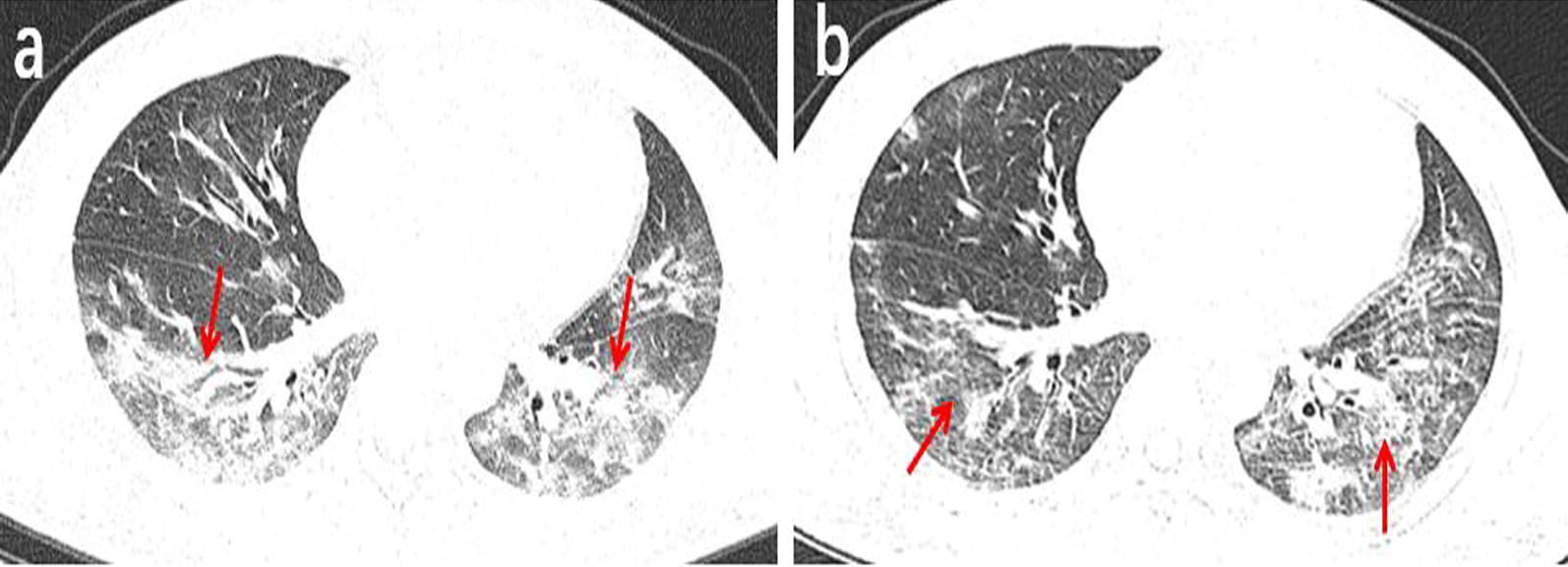
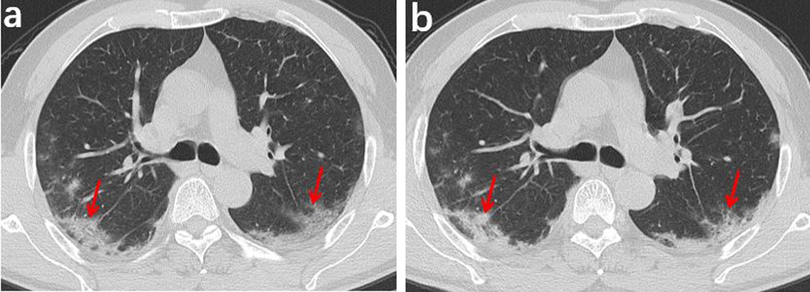
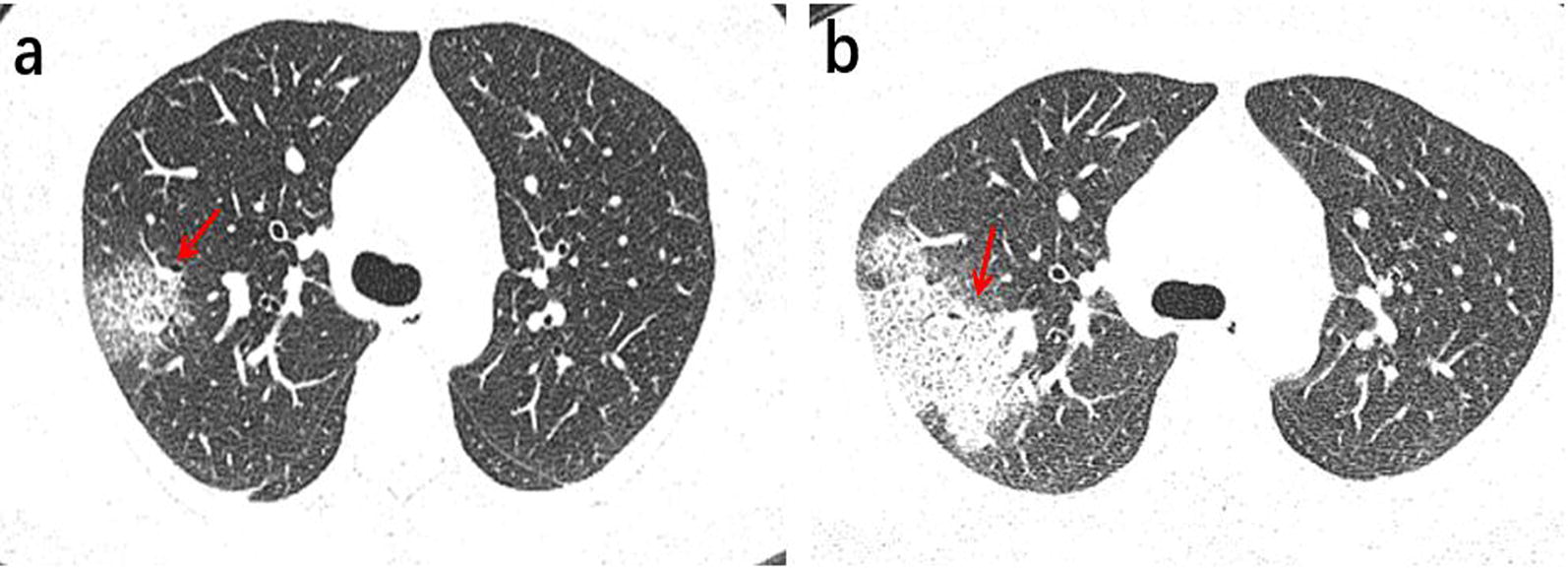
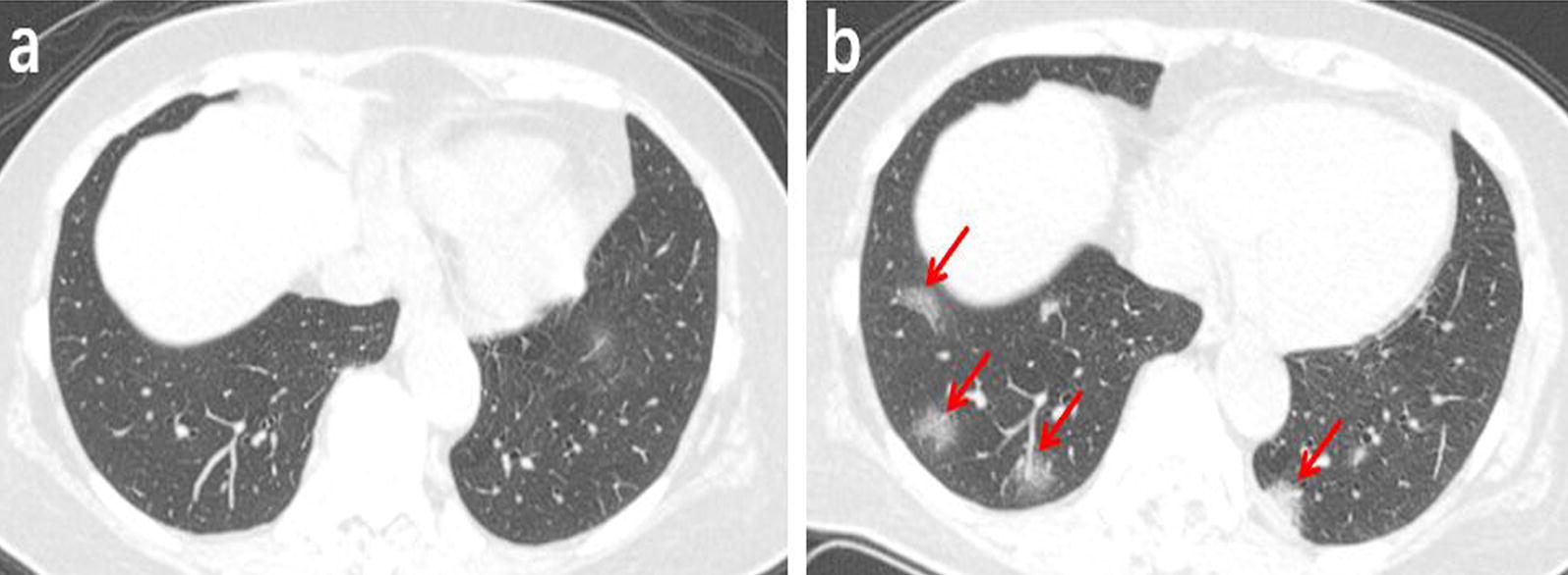
Results: A total of 100 (76%) patients had a history of close contact with people living in Wuhan, Hubei. The clinical manifestations of COVID-19 included cough, fever. Most of the lesions identified in chest CT images were multiple lesions of bilateral lungs, lesions were more localized in the peripheral lung, 109 (83%) patients had more than two lobes involved, 20 (15%) patients presented with patchy ground glass opacities, patchy ground glass opacities and consolidation of lesions co-existing in 61 (47%) cases. Complications such as pleural thickening, hydrothorax, pericardial effusion, and enlarged mediastinal lymph nodes were detected but only in rare cases. For the follow-up chest CT examinations (91 cases), We found 66 (73%) cases changed very quickly, with an average of 3.5 days, 25 cases (27%) presented absorbed lesions, progression was observed in 41 cases (46%), 25 (27%) cases showed no significant changes.
Conclusion: Chest CT plays an important role in diagnosing COVID-19. The imaging pattern of multifocal peripheral ground glass or mixed consolidation is highly suspicious of COVID-19, that can quickly change over a short period of time.
Keywords: Computed tomography; Coronavirus; Evolvement; Pneumonia; The chest.
Conflict of interest statement
The authors declare that they have no competing interests.
Figures
References
-
- Gorbalenya AE, Baker SC, Baric RS, de Groot RJ, Drosten C, Gulyaeva AA, Haagmans BL, Lauber C, Leontovich AM, Neuman BW, et al. Severe acute respiratory syndrome-related coronavirus-the species and its virusesa statement of the Coronavirus Study Group. BioRxiv. 2020 doi: 10.1101/2020.02.07.937862. - DOI
-
- Guan WJ, Ni ZY, Hu Y, Liang WH, Ou CQ, He JX, Liu L, Shan H, Lei CL, Hui DS, et al. Clinical characteristics of 2019 novel coronavirus infection in China. MedRxiv. 2020 doi: 10.1101/2020.02.06.20020974. - DOI
-
- Radiological Diagnosis of New Coronavirus Infected Pneumonitis: Expert Recommendation from the Chinese Society of Radiology (First edition), Chin J Radiol 2020,54(00): E001-E001.
Publication types
MeSH terms
Grants and funding
- cstc2018jszx-cyztzxX0017/the research about Chongqing Key technology and application demonstration of medical imaging depth intelligent diagnostic platform/International
- 2017MPRC-07/Nursery Talents of Army Medical University/International
- SWH2018QNWQ-04/Military medical innovation ability improvement plan of medical staff in the First Affiliated Hospital to Army Medical University/International
- 2016YFC0107101/National Key Research and Development Project/International
LinkOut - more resources
Full Text Sources
