. 2020 Apr 6;39(1):59.
doi: 10.1186/s13046-020-01553-7.

Cullin-7 (CUL7) is overexpressed in glioma cells and promotes tumorigenesis via NF-κB activation

Affiliations

Cullin-7 (CUL7) is overexpressed in glioma cells and promotes tumorigenesis via NF-κB activation

Jianye Xu et al. J Exp Clin Cancer Res. .

Abstract

Background: Cullin-7 (CUL7) is a member of the DOC domain-containing cullin family and is involved in the regulation of cell transformation. However, the clinical significance, potential mechanism and upstream regulators of CUL7 in malignant gliomas remain to be determined.

Methods: Expression level data and clinical information were obtained via the Cancer Genome Atlas (TCGA) database, the Chinese Glioma Genome Atlas (CGGA) database, immunohistochemistry (IHC) and western blot analysis. Gene set enrichment analysis (GSEA) was used to explore the potential molecular mechanisms of CUL7. RNA silencing was performed using siRNA or lentiviral constructs in U87MG and U251 glioma cell lines and GSC267 glioma stem cells. CUL7 overexpression was performed using the GV141-CUL7 plasmid construct. In addition, overexpression of miR-3940-5p was performed and validated by quantitative real-time PCR (qRT-PCR). Cells were characterized in vitro or in vivo to evaluate their molecular status, cell proliferation, invasion, and migration by Cell Counting Kit (CCK)-8, EdU, flow cytometry, colony formation, Transwell and 3D tumour spheroid invasion assays. Coimmunoprecipitation (co-IP) and western blotting were performed to test the mechanisms of activation of the NF-κB signalling pathway.

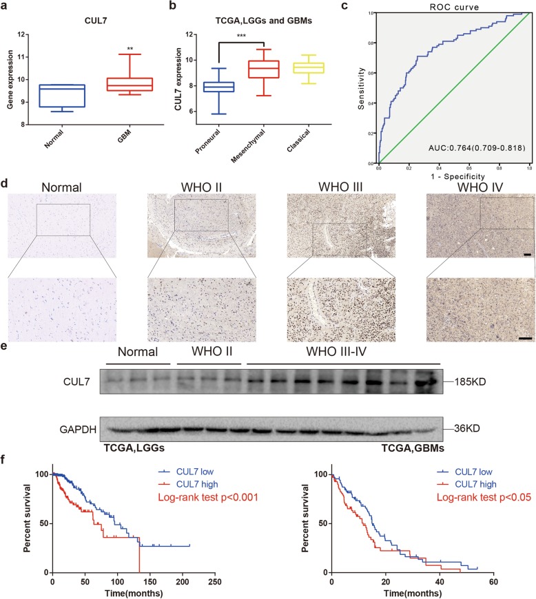
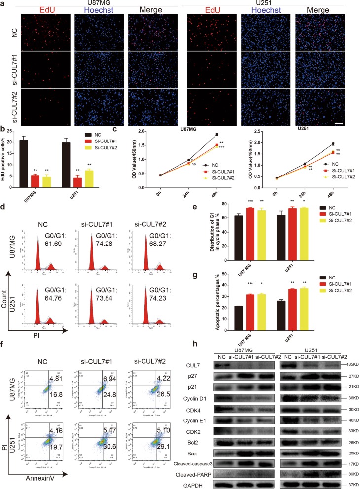
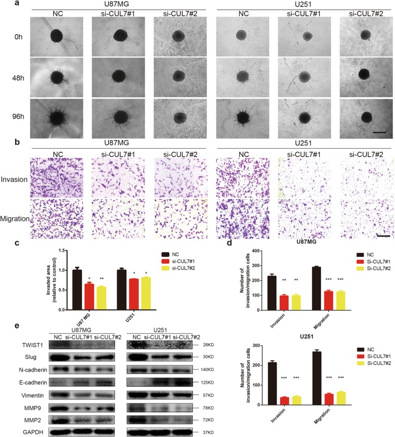
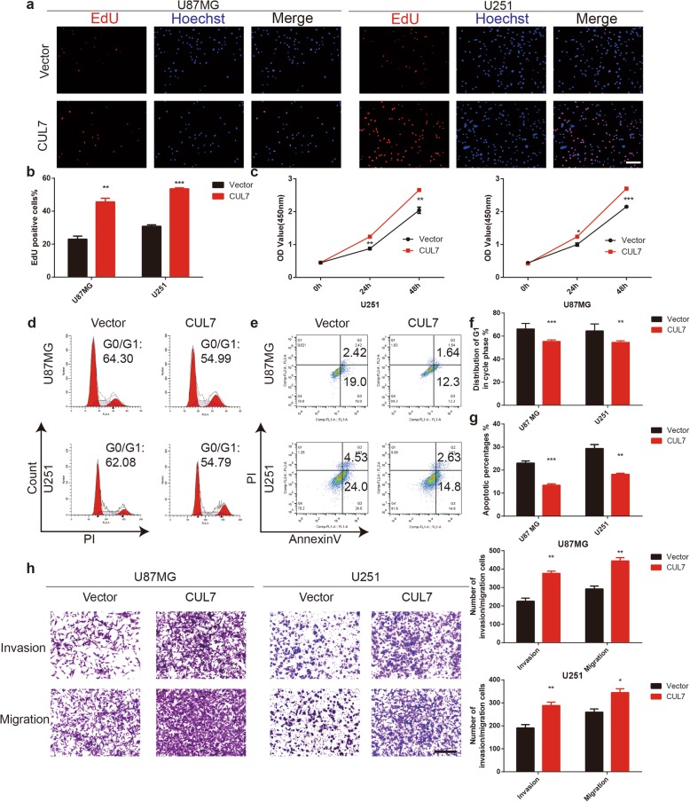
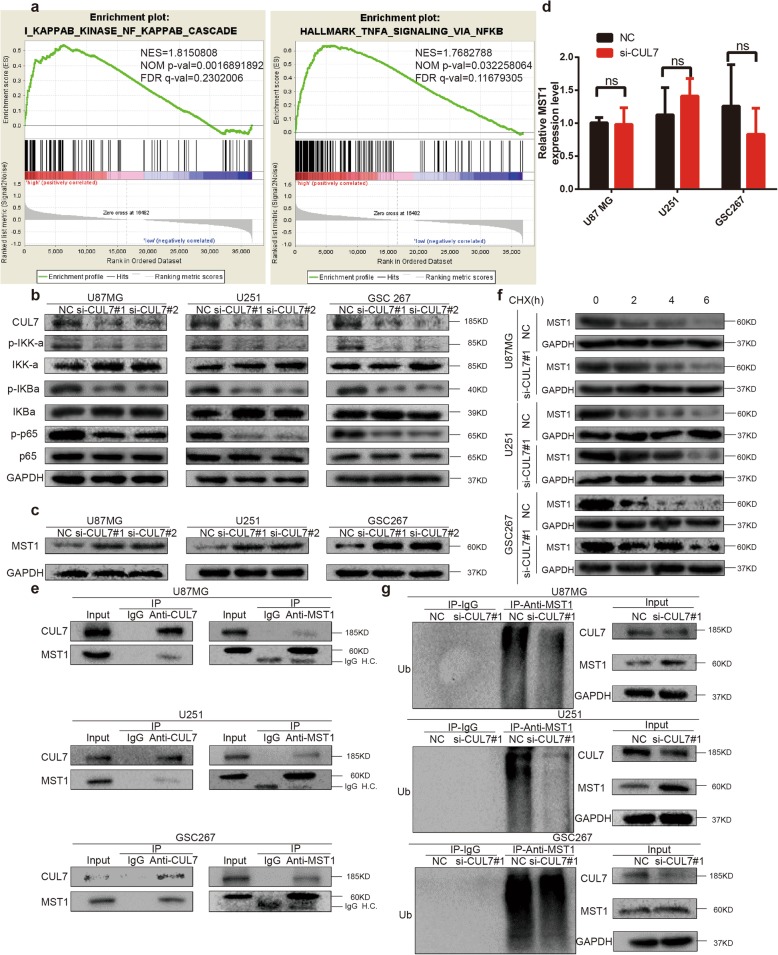
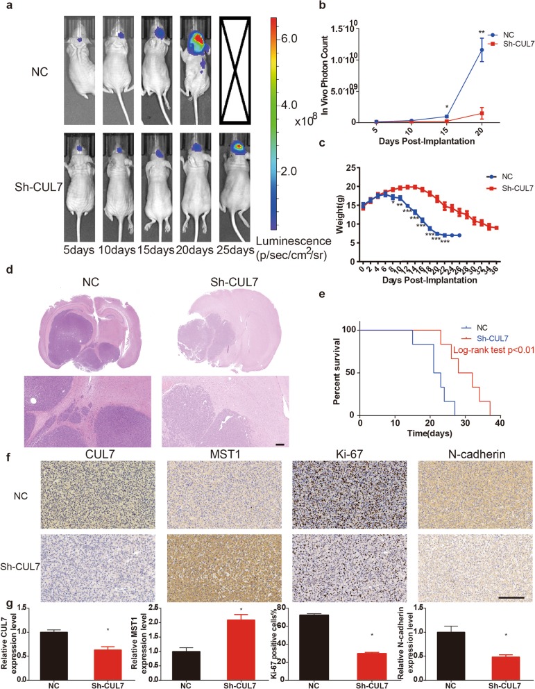
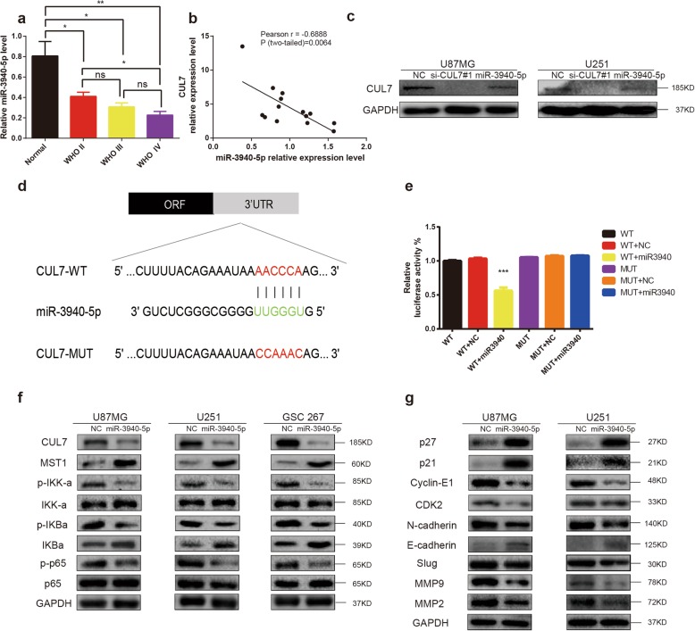
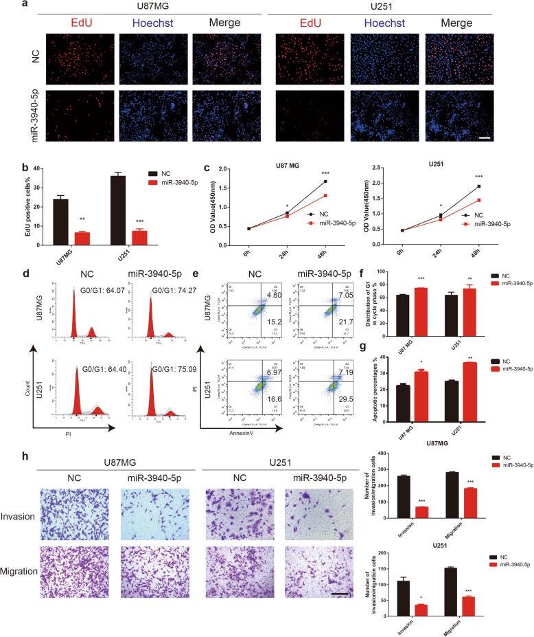
Results: High CUL7 expression was associated with a high tumour grade, a mesenchymal molecular glioma subtype and a poor prognosis in patients. Gene silencing of CUL7 in U87MG and U251 cells significantly inhibited tumour growth, invasion and migration in vitro and in vivo. Western blot analysis revealed that cyclin-dependent kinase inhibitors and epithelial-mesenchymal transition (EMT) molecular markers changed under CUL7 silencing conditions. In contrast, CUL7 overexpression promoted tumour growth, invasion and migration. Gene set enrichment analysis (GSEA) and western blot analysis revealed that CUL7 was positively associated with the NF-κB pathway. Moreover, with coimmunoprecipitation assays, we discovered that CUL7 physically associated with MST1, which further led to ubiquitin-mediated MST1 protein degradation, which promoted activation of the NF-κB signalling pathway. Finally, CUL7 was found to be downregulated by miR-3940-5p, which suppressed the development of gliomas.

Conclusions: Our findings indicate that CUL7 plays a significant role in promoting tumorigenesis via NF-κB activation and that it can be negatively regulated by miR-3940-5p in human gliomas. Furthermore, CUL7 might be a candidate molecular target for the treatment of glioma.

Keywords: CUL7; Glioma; MST1; NF-κB; miR-3940-5p.


Conflict of interest statement

The authors declare that they have no competing interests.

Figures

Fig. 1
Fig. 1
Expression of CUL7 is associated with tumor grade and patient survival in gliomas. a Quantification of CUL7 mRNA expression levels in GBMs and normal brain tissues in TCGA. b Quantification of gliomas subtype-specific CUL7 expression in TCGA. Log2-transformed expression of CUL7 mRNA levels are listed on the Y-axis. Error bars represents the SEM. c ROC curve showing sensitivity of CUL7 as a marker to discriminate between mesenchymal subtype and non-mesenchymal subtype glioma patients. d Representative images of IHC staining for CUL7 in different grade gliomas and normal brain tissues (scale bar = 100 μm). e Western blot analysis of CUL7 levels in lysates from different grades of glioma tissues (WHO grades II–IV) and normal brain tissues. f Kaplan–Meier survival analysis for glioma patients with high CUL7 expression and low CUL7 expression in LGGs (n = 457) or GBMs (n = 141) in TCGA database. The cut-off level was set at the median value of the CUL7 levels. *P < 0.05; **P < 0.01; ***P < 0.001
Fig. 2
Fig. 2
CUL7 knockdown inhibits cell proliferation and induces cell cycle arrest and cell apoptosis. U87MG and U251 cells transfected with CUL7 siRNA or controls and characterized in the following assays: (a, b) EdU performed 48 h after transfection (scale bar = 100 μm); (c) growth curve based on OD450 using the CCK-8 assay; (d, e) cell cycle profiles determined from PI staining in flow cytometry; (f, g) % apoptosis as determined with Annexin V-FITC antibody and PI staining in flow cytometry. (h) Western blot to detect expression levels of the known cell cycle and apoptosis regulatory factors indicated. GAPDH was used as a loading control. Data are represented as the mean ± SEM from three independent experiments. *P < 0.05; **P < 0.01; ***P < 0.001, relative to control.NC: non-silencing siRNA; si-CUL7: siRNAs targeting CUL7
Fig. 3
Fig. 3
CUL7 knockdown decreases invasive and migrative ability of glioma cells. a Representative images of invaded spheroids in 3D invasion assay for U87MG and U251 transfected with CUL7 and control siRNAs evaluated at 48 h and 96 h are shown (Scale bar = 200 μm). b Representative images of Transwell migration and invasion assays performed in U87MG and U251 transfected NC and si–CUL7 cells (Scale bar = 200 μm). c The area covered by invading cells quantitated after 96 h. d Graphic representation of migrated and invaded cells counts from Transwell assay. Data are represented as the mean ± SEM from three independent experiments. *P < 0.05; **P < 0.01; ***P < 0.001, relative to control. e Western blot for protein levels of EMT components in lysates (20 μg) from U87MG and U251 cells transfected with siRNA against CUL7 and controls. GAPDH was used as a loading control. NC: non-silencing siRNA; si-CUL7: siRNAs targeting CUL7
Fig. 4
Fig. 4
CUL7 overexpression promotes proliferation, invasion and migration of glioma cells. U87MG and U251 cells transfected with GV141-CUL7s or Vectors and characterized in the following assays: (a, b) EdU performed 48 h after transfection (scale bar = 100 μm); (c) growth curve based on OD450 using the CCK-8 assay; (d, f) cell cycle profiles determined from PI staining in flow cytometry (e, g) % apoptosis as determined with Annexin V-FITC antibody and PI staining in flow cytometry. h Representative images of Transwell migration and invasion assays performed in U87MG and U251 cells. Graphic representation of migrated and invaded cells counts from Transwell assay. Data are represented as the mean ± SEM from three independent experiments (Scale bar = 200 μm). *P < 0.05; **P < 0.01; ***P < 0.001, relative to control. Vector: GV141-empty; CUL7: GV141-CUL7
Fig. 5
Fig. 5
CUL7 leads to ubiquitin-mediated MST1 protein degradation and promotes activation of the NF-κB pathway. a GSEA highlighting positive association of increased CUL7 expression levels with NF-κB signal pathway. NES = normalized enrichment score; NOM = nominal FDR = false discovery rate. b Western blot for protein levels of NF-κB pathway components in lysates (20 μg) from U87MG, U251 and GSC267 cells transfected with siRNA against CUL7 and controls. GAPDH was used as a loading control. c Western blot for protein levels of MST1 in lysates (20 μg) from U87MG, U251 and GSC267 cells transfected with siRNA against CUL7 and controls. d qRT-PCR analysis of MST1 in U87MG, U251 and GSC267 cells. Expression is normalized to GAPDH mRNA. Data are represented as the mean ± SEM. e Western blot analysis of co-precipitating proteins in IPs performed using anti-CUL7 or -MST1 antibody on lysates prepared from U87MG, U251 and GSC267-NC, si-CUL7 cells. f Western blot analysis of MST1 protein in modified U87MG, U251 and GSC267 cells treated with CHX (25 μg/mL) for the indicated time. g Western blot analysis of MST1 IPs performed on lysates prepared from U87MG, U251 and GSC267 cells treated with MG132 (20 μM) for 8 h to examine endogenous MST1 ubiquitination. NC: non-silencing siRNA; si-CUL7: siRNAs targeting CUL7
Fig. 6
Fig. 6
CUL7 silencing inhibits tumorigenesis in vivo. a, b Bioluminescence imaging showed the tumor size as days elapsed. c Weights of two groups of mice were measured every two days. d H&E staining of sections from mouse brains with U87MG control or sh-CUL7 xenografts at 15 days after implantation with 3 × 10⁶ cells. (scale bar = 200 μm) e Survival analysis for animals implanted with U87MG sh-CUL7 or control cells (P < 0.01 by log-rank test; n control = 6, n sh-CUL7 = 6). f, g IHC for CUL7, MST1, Ki-67 and N-cadherin in sections from indicated xenografts (scale bar = 100 μm). Data are represented as the mean ± SEM
Fig. 7
Fig. 7
CUL7 is a direct target of miR-3940-5p in glioma cells. a qRT-PCR analysis validated the lower expression of miR-3940-5p in glioma tissues. b Pearson r correlation was used to analyze the relationship between CUL7 and miR-3940-5p in Qilu hospital patient samples. (n = 14). c Overexpression of miR-3940-5p markedly suppressed the protein levels of CUL7 in glioma cells. d miR-3940-5p and its putative binding sequence in the wild-type (WT) and mutant (MUT) 3′-UTR of CUL7. e Overexpression of miR-3940-5p significantly decreased the luciferase activity that carried wild-type (WT) but not mutant type (MUT) 3′-UTR of CUL7 in glioma cells. f Western blot to detect expression levels of the MST1 and markers of activation of NF-κB pathway. GAPDH was used as a loading control. g Western blot for protein levels of cell cycle regulatory factors and EMT components in lysates (20 μg) from U87MG and U251 cells transfected with miR-3940-5p mimics and controls. GAPDH was used as a loading control. *P < 0.05; **P < 0.01; ***P < 0.001; NC: negative control RNA; si-CUL7: siRNAs targeting CUL7; miR-3940-5p: miR-3940-5p mimics
Fig. 8
Fig. 8
MiR-3940-5p inhibits cell proliferation, migration and invasion of glioma. U87MG and U251 cells transfected with miR-3940-5p mimics or controls and characterized in the following assays: (a, b) EdU performed 48 h after transfection (scale bar = 100 μm); (c) growth curve based on OD450 using the CCK-8 assay; (d, f) cell cycle profiles determined from PI staining in flow cytometry (e, g) % apoptosis as determined with Annexin V-FITC antibody and PI staining in flow cytometry. (h) Representative images of transwell migration and invasion assays performed in U87MG and U251 transfected NC and miR-3940-5p cells. Graphic representation of migrated and invaded cells counts from transwell assay (Scale bar = 200 μm). Data are represented as the mean ± SEM from three independent experiments. *P < 0.05; **P < 0.01; ***P < 0.001, relative to control. NC: negative control RNA; miR-3940-5p: miR-3940-5p mimics

Similar articles

Cited by

References

    1. Furnari FB, Fenton T, Bachoo RM, Mukasa A, Stommel JM, Stegh A, et al. Malignant astrocytic glioma: genetics, biology, and paths to treatment. Genes Dev. 2007;21(21):2683–2710. doi: 10.1101/gad.1596707. - DOI - PubMed
    1. Lapointe S, Perry A, Butowski NA. Primary brain tumours in adults. Lancet. 2018;392(10145):432–446. doi: 10.1016/S0140-6736(18)30990-5. - DOI - PubMed
    1. Wen PY, Kesari S. Malignant Gliomas in adults. N Engl J Med. 2008;359(5):492–507. doi: 10.1056/NEJMra0708126. - DOI - PubMed
    1. Torre LA, Bray F, Siegel RL, Ferlay J, Lortet-Tieulent J, Jemal A. Global cancer statistics, 2012. CA Cancer J Clin. 2015;65(2):87–108. doi: 10.3322/caac.21262. - DOI - PubMed
    1. Wang Q, Hu B, Hu X, Kim H, Squatrito M, Scarpace L, et al. Tumor evolution of Glioma-intrinsic gene expression subtypes associates with immunological changes in the microenvironment. Cancer Cell. 2018;33(1):152. doi: 10.1016/j.ccell.2017.12.012. - DOI - PMC - PubMed

MeSH terms