Review
. 2020 Apr 6;13(1):170.
doi: 10.1186/s13071-020-04044-y.

Characteristic features of the SERA multigene family in the malaria parasite

Affiliations
Review

Characteristic features of the SERA multigene family in the malaria parasite

Nobuko Arisue et al. Parasit Vectors. .

Abstract

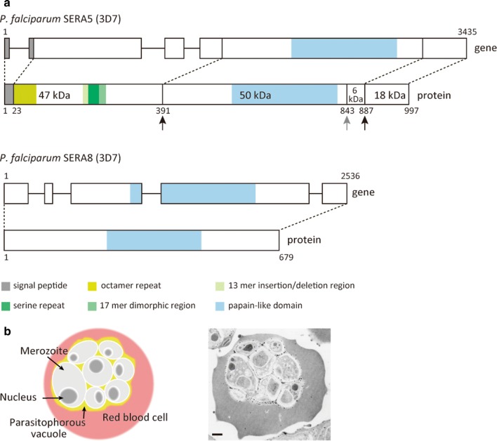
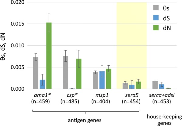
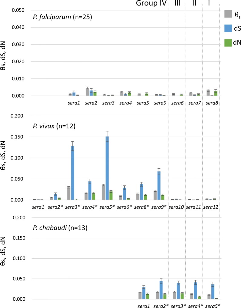
Serine repeat antigen (SERA) is conserved among species of the genus Plasmodium. Sera genes form a multigene family and are generally tandemly clustered on a single chromosome. Although all Plasmodium species encode multiple sera genes, the number varies between species. Among species, the members share similar sequences and gene organization. SERA possess a central papain-like cysteine protease domain, however, in some members, the active site cysteine residue is substituted with a serine. Recent studies implicate this gene family in a number of aspects in parasite biology and induction of protective immune response. This review summarizes the current understanding on this important gene family in several Plasmodium species. The Plasmodium falciparum (Pf)-sera family, for example, consists of nine gene members. Unlike other multigene families in Plasmodium species, Pf-sera genes do not exhibit antigenic variation. Pf-sera5 nucleotide diversity is also low. Moreover, although Pf-sera5 is highly transcribed during the blood stage of malaria infection, and a large amount is released into the host blood following schizont rupture, in malaria endemic countries the sero-positive rates for Pf-SERA5 are low, likely due to Pf-SERA5 binding of host proteins to avoid immune recognition. As an antigen, the N-terminal 47 kDa domain of Pf-SERA5 is a promising vaccine candidate currently undergoing clinical trials. Pf-SERA5 and Pf-SERA6, as well as P. berghei (Pb)-SERA3, and Pb-SERA5, have been investigated for their roles in parasite egress. Two P. yoelii SERA, which have a serine residue at the protease active center, are implicated in parasite virulence. Overall, these studies provide insight that during the evolution of the Plasmodium parasite, the sera gene family members have increased by gene duplication, and acquired various functions that enable the parasite to survive and successfully maintain infection in the host.

Keywords: Function; Gene family; Plasmodium; Polymorphism; SERA.


Conflict of interest statement

The authors declare that they have no competing interests.

Figures

Fig. 1
Fig. 1
Plasmodium sera gene family. a The organization of Plasmodium sera genes and their characteristic gene and protein features. Plasmodium sera genes (denoted as circles) are clustered in tandem and are categorized into four groups based on genetic background. In terms of protein expression, i: Group I SERA is expressed in the mosquito stage and Group II to IV are expressed in the host blood stage. In terms of the protease motif, ii: SERA members containing cysteine residues in the protease active center belong to Groups I to III, while those with a substituted serine residue belong to Group IV. The number of Group IV SERA genes can vary from 1 to 11. b The number of sera genes identified in each group. For Group IV sera and two Group I sera, the number of genes not in tandem cluster is shown in parenthesis. Plasmodium species with humans as a natural host are shown in red. The number of Group IV sera genes in P. adleri is currently unresolved
Fig. 2
Fig. 2
Phylogenetic tree of sera genes inferred by the maximum likelihood (ML) method. The sequences of Plasmodium sera genes were translated to amino acid sequences and aligned by using MAFFT version 7.409 [58]. Site selection for tree inference was done with the Gblocks option, with a more stringent selection criteria implemented in SeaView version 4.7 [59]. Trees were inferred under the JTT+Г+I model. MEGA X [60] was used for the analysis. Bootstrap proportions from the ML methods with 100 replications are shown. a 300 amino acid positions from 195 Group I to IV sera genes were used for tree inference. The tree with the highest log likelihood (−li = 28137.0426) is shown. A discrete Г distribution was used to model evolutionary rate differences among sites (4 categories, +Г, parameter = 1.1871). The rate variation model allowed for some sites to be evolutionarily invariable (+I, 9.2557% sites). b 424 amino acid positions from 173 Group II to IV sera genes were used for tree inference. The log likelihood of the tree is − 37969.9. A discrete Г distribution was used to model evolutionary rate differences among sites (4 categories, +Г, parameter = 1.2356). The rate variation model allowed for some sites to be evolutionarily invariable (+I, 5.0708% sites)
Fig. 3
Fig. 3
Structure and localization of SERA. a Schematic representation of the Plasmodium falciparum SERA5 and SERA8 genes and proteins; P. falciparum 3D7 strain was used as a reference for the illustration. The gene IDs in PlasmoDB [18] were PF3D7_0207600 and PF3D7_0207300, for Pf-SERA5 and Pf-SERA8, respectively. Amino acid positions for the Pf-SERA5 fragments are: 47 kDa corresponding to 23–390 aa; 50 kDa: 391–842 aa; 6 kDa: 843–886 aa; and 18 kDa: 887–997 aa. Black arrows indicate the PfSUB1 cleavage sites. The 50 kDa + 6 kDa fragment is further processed by an unknown protease (gray arrow). b Localization of Pf-SERA5 in the parasitophorous vacuole. Left image shows an infected red blood cell with schizont stage malaria parasites inside the parasitophorous vacuole. Right image, N-terminal antisera was used to localize Pf-SERA5 within a schizont parasitized red blood cell. Viewed under JEM-1230 transmission electron microscope (18,000× magnification). SERA5 conjugated gold particles were observed inside the parasitophorous vacuole. Scale-bar: b, 500 nm
Fig. 4
Fig. 4
Plasmodium life-cycle and SERA function. The parasite life-cycle in the mosquito vector and human host. The colored circles indicate where SERA function is proposed by current studies
Fig. 5
Fig. 5
Polymorphism of P. falciparum antigen genes and housekeeping genes. The amount of nucleotide diversity expressed by the standardized number of polymorphic sites per site (θs), the numbers of synonymous substitutions per synonymous site (dS), and non-synonymous substitutions per non-synonymous site (dN). Data were taken from Tanabe et al. [45, 47]. The asterisk denotes a significant difference between dS and dN (P < 0.01). Abbreviations: n, number of sequences analyzed; ama1, apical membrane protein 1; csp, circumsporozoite protein; msp1, merozoite surface protein 1; sera5, serine repeat antigen 5; serca, P-type Ca2+-ATPase; adsl, adenylosuccinate lyase
Fig. 6
Fig. 6
Sequence diversities (θs), dS, dN in P. falciparum, P. vivax, and P. chabaudi sera genes. The amount of nucleotide diversity as expressed by the standardized number of polymorphic sites per site (θs) were calculated using DnaSP v5.10.01 [61]. The numbers of synonymous substitutions per synonymous site (dS) and of nonsynonymous substitutions per nonsynonymous site (dN) were calculated using MEGA X [60]. As some strains of Pv-sera7 contain stop codons in the predicted open reading frame, sequences of Pv-sera7 were omitted from analysis. The asterisk denotes significant difference between dS and dN (P < 0.01)

References

    1. WHO. World malaria report 2019. Geneva: World Health Organisation; 2019. https://www.who.int/news-room/feature-stories/detail/world-malaria-repor.... Accessed 19 Dec 2019.
    1. Bzik DJ, Li WB, Horii T, Inselburg J. Amino acid sequence of the serine repeat antigen (SERA) of Plasmodium falciparum determined from cloned cDNA. Mol Biochem Parasitol. 1988;30:279–288. doi: 10.1016/0166-6851(88)90097-7. - DOI - PubMed
    1. Gardner MJ, Tettelin H, Carucci DJ, Cummings LM, Aravind L, Koonin EV, et al. Chromosome 2 sequence of the human malaria parasite Plasmodium falciparum. Science. 1998;282:1126–1132. doi: 10.1126/science.282.5391.1126. - DOI - PubMed
    1. Hall N, Pain A, Berriman M, Churcher C, Harris B, Harris D, et al. Sequence of Plasmodium falciparum chromosomes 1, 3–9 and 13. Nature. 2002;419:527–531. doi: 10.1038/nature01095. - DOI - PubMed
    1. Le Roch KG, Zhou Y, Blair PL, Grainger M, Moch JK, Haynes JD, et al. Discovery of gene function by expression profiling of the malaria parasite life cycle. Science. 2003;301:1503–1508. doi: 10.1126/science.1087025. - DOI - PubMed

Substances